Copyright
©The Author(s) 2022.
World J Clin Cases. Nov 6, 2022; 10(31): 11597-11606
Published online Nov 6, 2022. doi: 10.12998/wjcc.v10.i31.11597
Published online Nov 6, 2022. doi: 10.12998/wjcc.v10.i31.11597
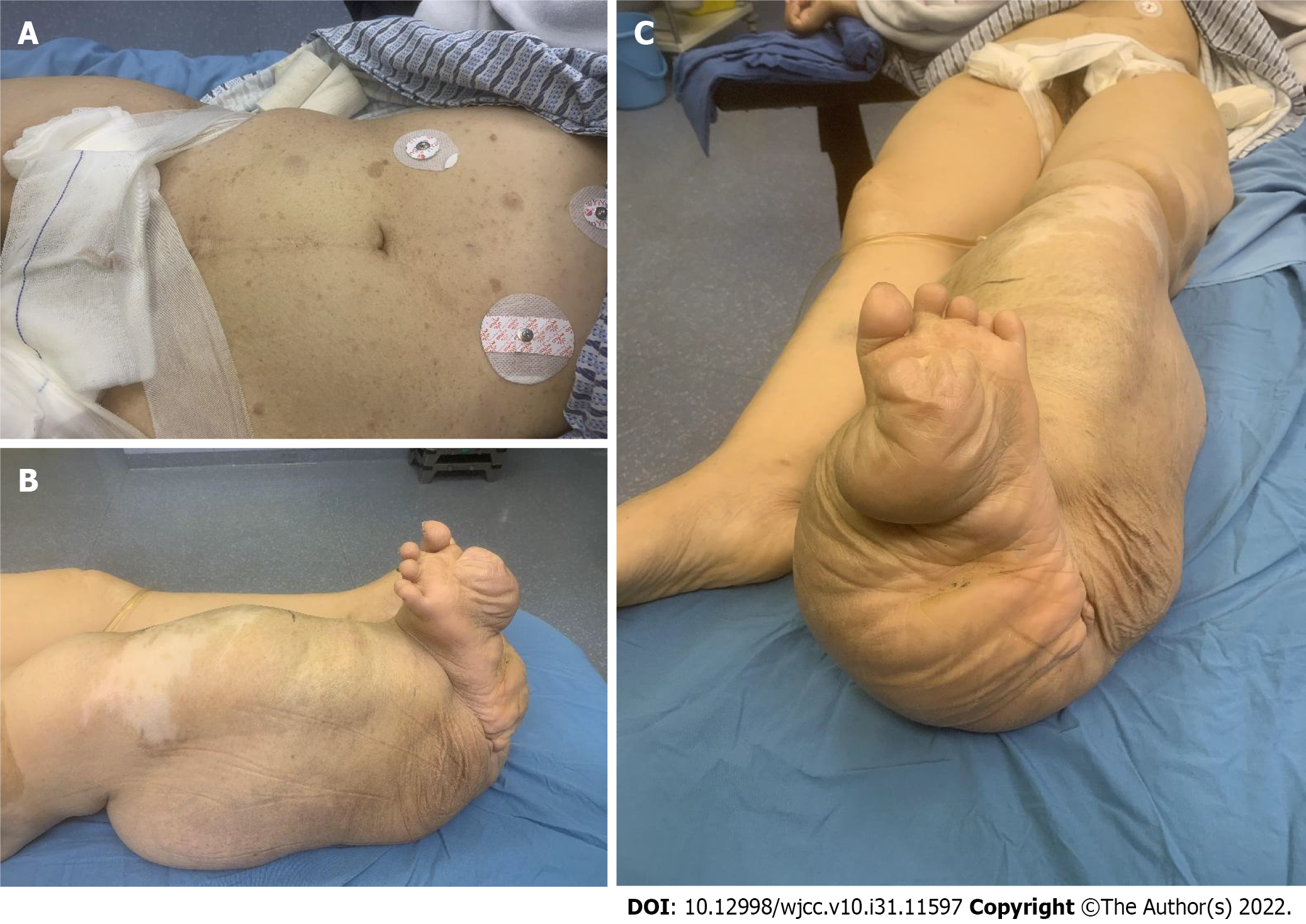
Figure 1 Presentation of the typical signs by the patient.
A: Multiple visible areas of dark-brown pigmentation on the abdomen; B and C: Right lower limb presenting as elephantiasis.
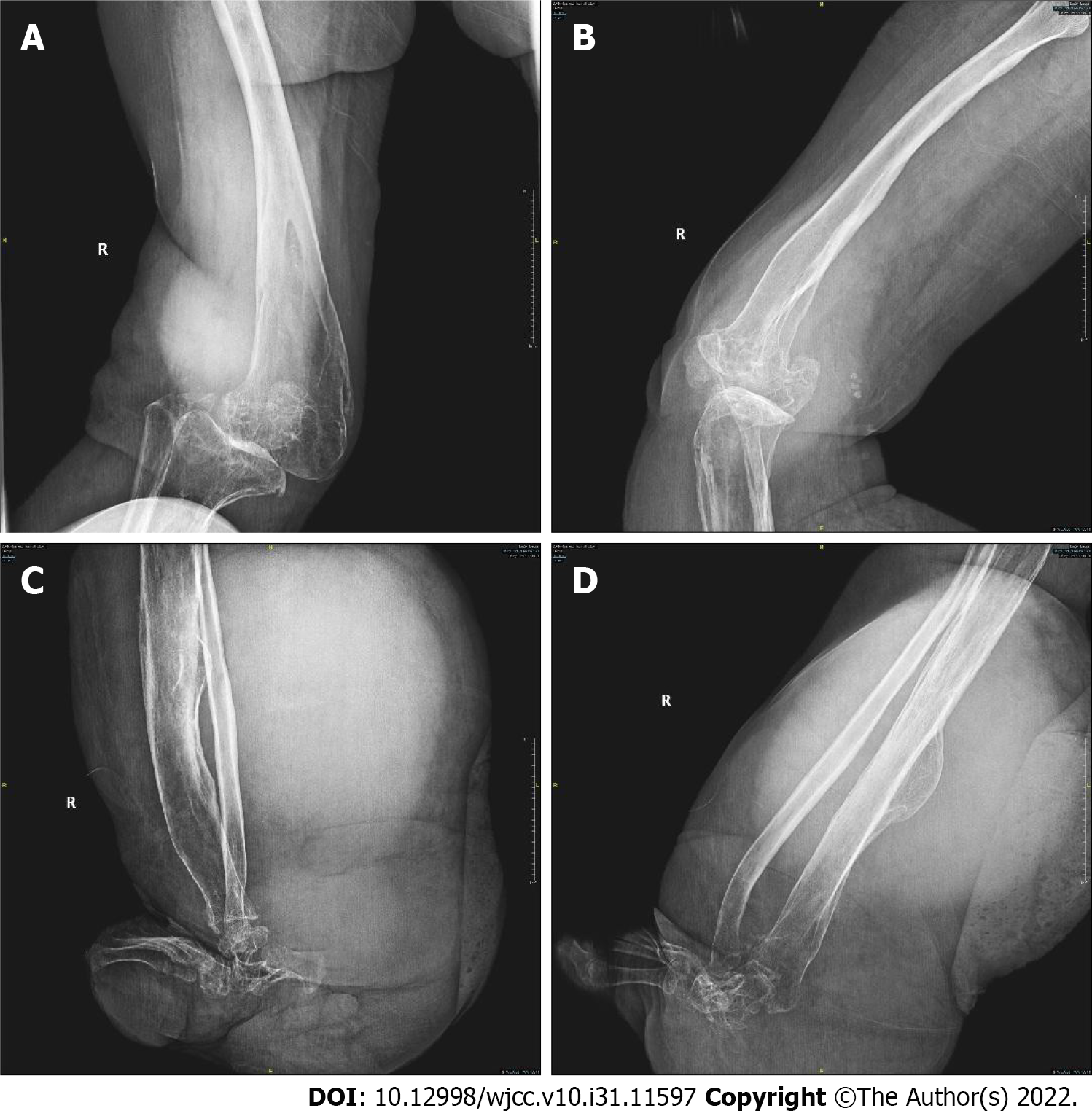
Figure 2 Preoperative imaging findings.
A and B: Growth disturbance at the distal femur and degenerative changes in the right knee joint; C and D: Bone morphological changes in the lower segment of the right tibia.
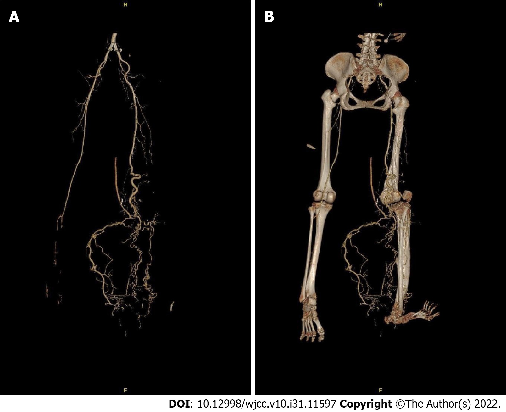
Figure 3 Preoperative computed tomographic angiography findings.
A: Computed tomographic angiography examination showed the following: the right popliteal and posterior tibial arteries were thickened and tortuous, multiple collateral small blood vessels were formed, and the superficial veins of the right calf were increased and thickened; B: 3D reconstruction of the right lower extremity skeleton revealed skeletal changes in the right lower extremity with growth disturbance and right knee subluxation.
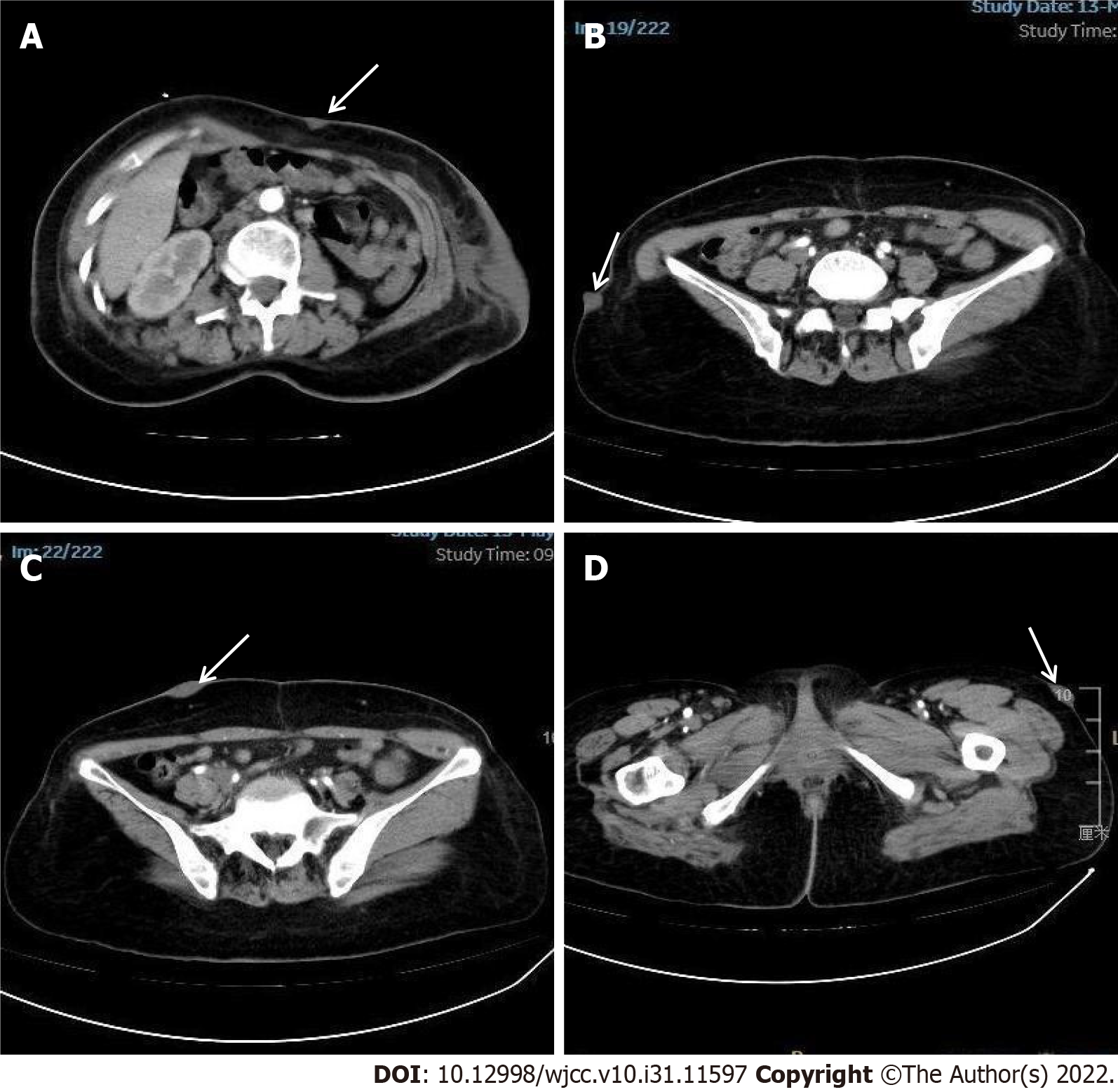
Figure 4 The soft tissue window of abdominal computed tomography.
Multiple nodules on the epidermis of the abdomen growing inward and outward (white arrow). A: Front of abdomen; B: Outside of right iliac; C: Anterior of right iliac; D: Outside of left hip.
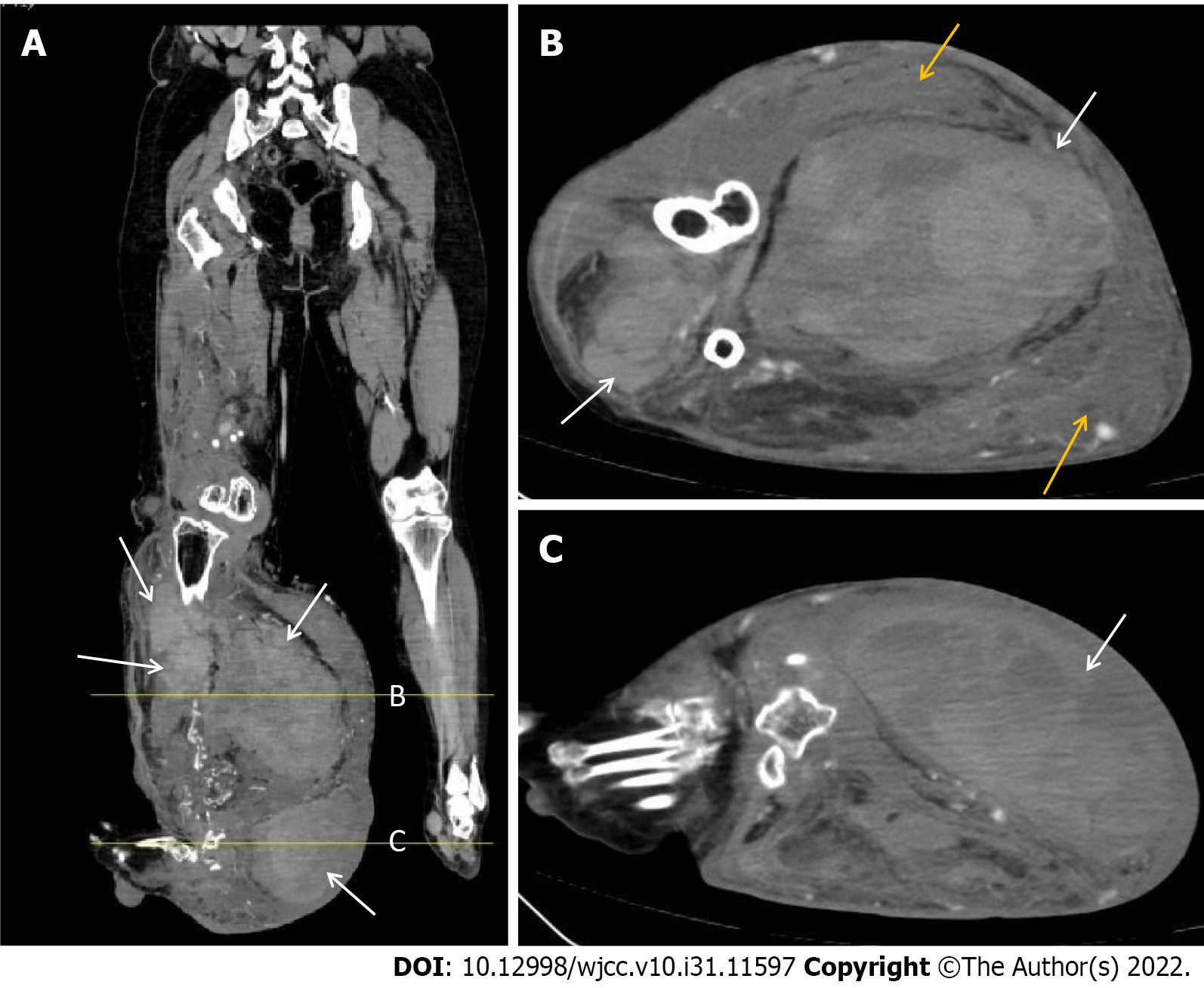
Figure 5 The soft tissue window of the right lower extremity computed tomography.
A: Sagittal view showed multiple large masses in the front and back of the right calf (white arrow, Lines A and B represent Figures A and B, respectively); B and C: Axial view showed multiple large masses in the front and back of the right calf and a subcutaneous hematoma behind the right calf (yellow arrow).
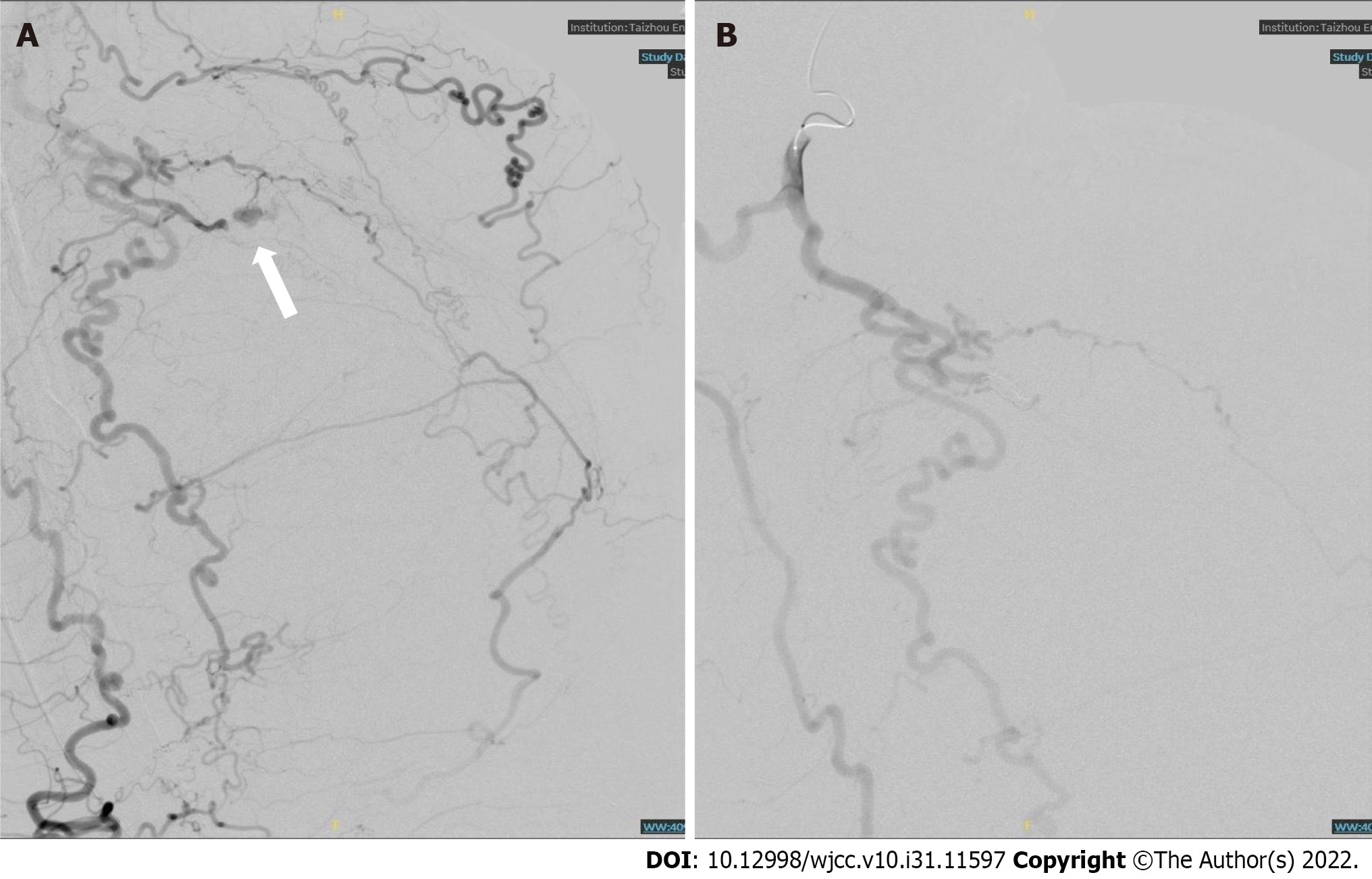
Figure 6 Right lower limb digital subtraction angiography and vascular embolization.
A: Digital subtraction angiography showed tortuous right popliteal and tibiofibular arteries and disordered tumor-like blood vessels. Contrast medium extravasation was seen in the local arterial branches in the right calf (white arrow); B: After vascular embolization, no contrast medium extravasation was found in the second angiography.
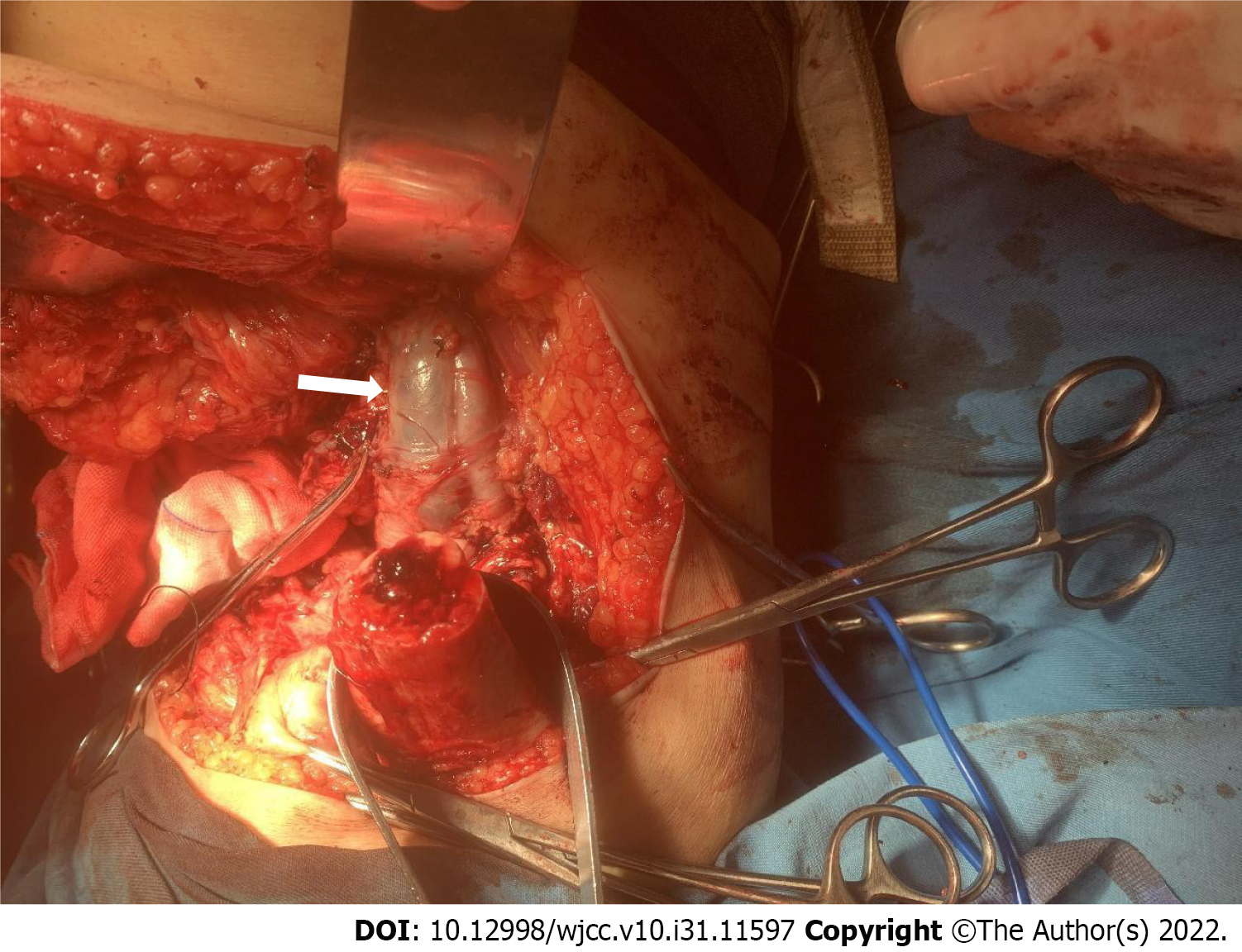
Figure 7 During the amputation operation, the femoral artery and vein were obviously thickened and tortuous.
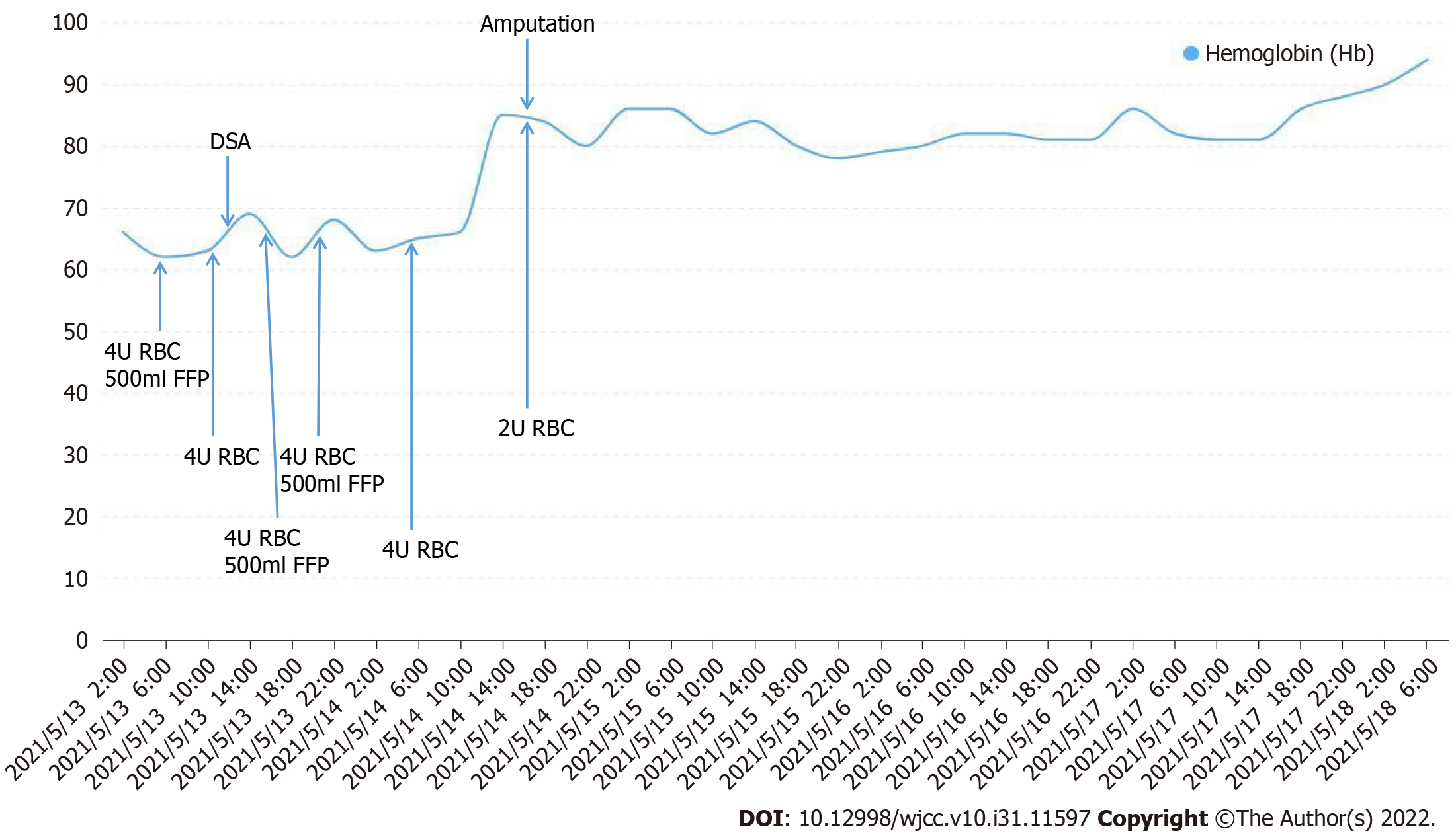
Figure 8 The trends of hemoglobin levels showed that after digital subtraction angiography and vascular embolization, the anemia and right lower limb swelling and tenderness did not improve.
After amputation, the patient's hemoglobin level improved significantly without blood transfusion. DSA: Digital subtraction angiography; RBCs: Red blood cells; FFP: Fresh frozen plasma.
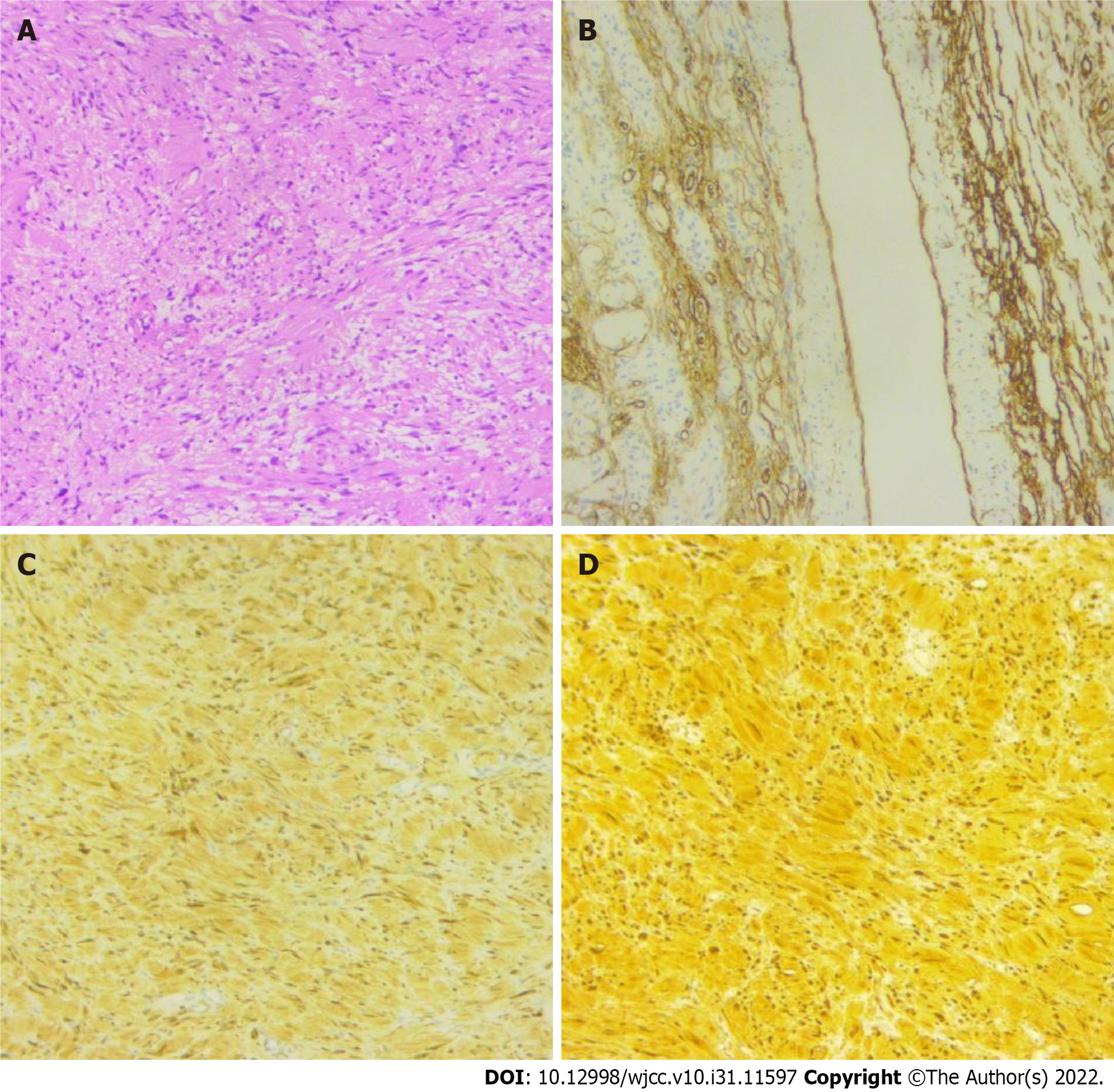
Figure 9 Postoperative pathology.
A: Tumor cells were spindle-shaped, arranged in fascicles and spirals, and mitoses were rare; B: Blood vessels were dilated and marked by CD34; C: Tumor cells were positive for Bcl-2 protein; D: Tumor cells were strongly positive for S-100 protein.
- Citation: Shen LP, Jin G, Zhu RT, Jiang HT. Hemorrhagic shock due to ruptured lower limb vascular malformation in a neurofibromatosis type 1 patient: A case report. World J Clin Cases 2022; 10(31): 11597-11606
- URL: https://www.wjgnet.com/2307-8960/full/v10/i31/11597.htm
- DOI: https://dx.doi.org/10.12998/wjcc.v10.i31.11597


