Copyright
©The Author(s) 2022.
World J Clin Cases. Nov 6, 2022; 10(31): 11590-11596
Published online Nov 6, 2022. doi: 10.12998/wjcc.v10.i31.11590
Published online Nov 6, 2022. doi: 10.12998/wjcc.v10.i31.11590
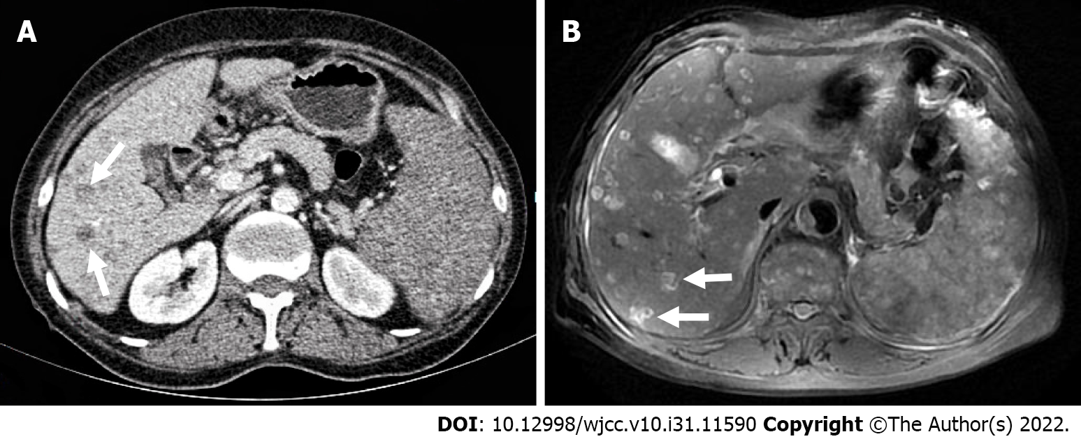
Figure 1 Contrast-enhanced computed tomography and contrast-enhanced magnetic resonance imaging reveal multiple low-density lesions in the liver with slightly low enhancement.
A: Contrast-enhanced computed tomography; B: Contrast-enhanced magnetic resonance imaging.
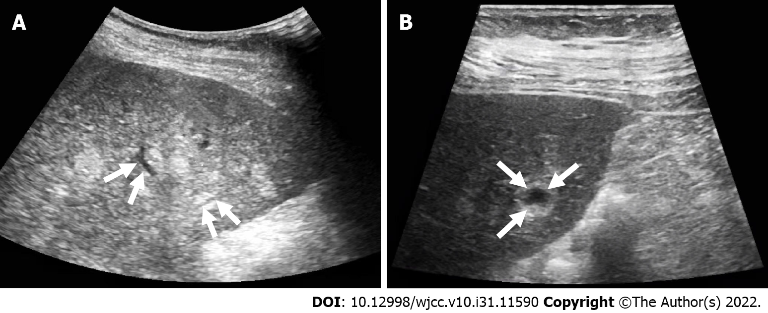
Figure 2 Conventional ultrasound.
A and B: Multiple intrahepatic hyperechoic nodules with unclear boundaries, loose structures, vascular-like structures (A) and anechoic areas (B).
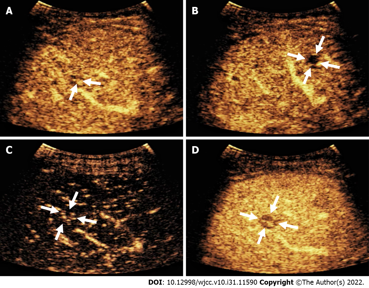
Figure 3 Contrast-enhanced ultrasound reveals nodular peripheral enhancement in the arterial and portal phases and low-enhancement and non-enhanced areas in the late phase.
A: Nodular peripheral enhancement; B: Non-enhanced area; C: The arterial phase showed low enhancement at 13 s; D: The portal vein was cleared at 58 s.
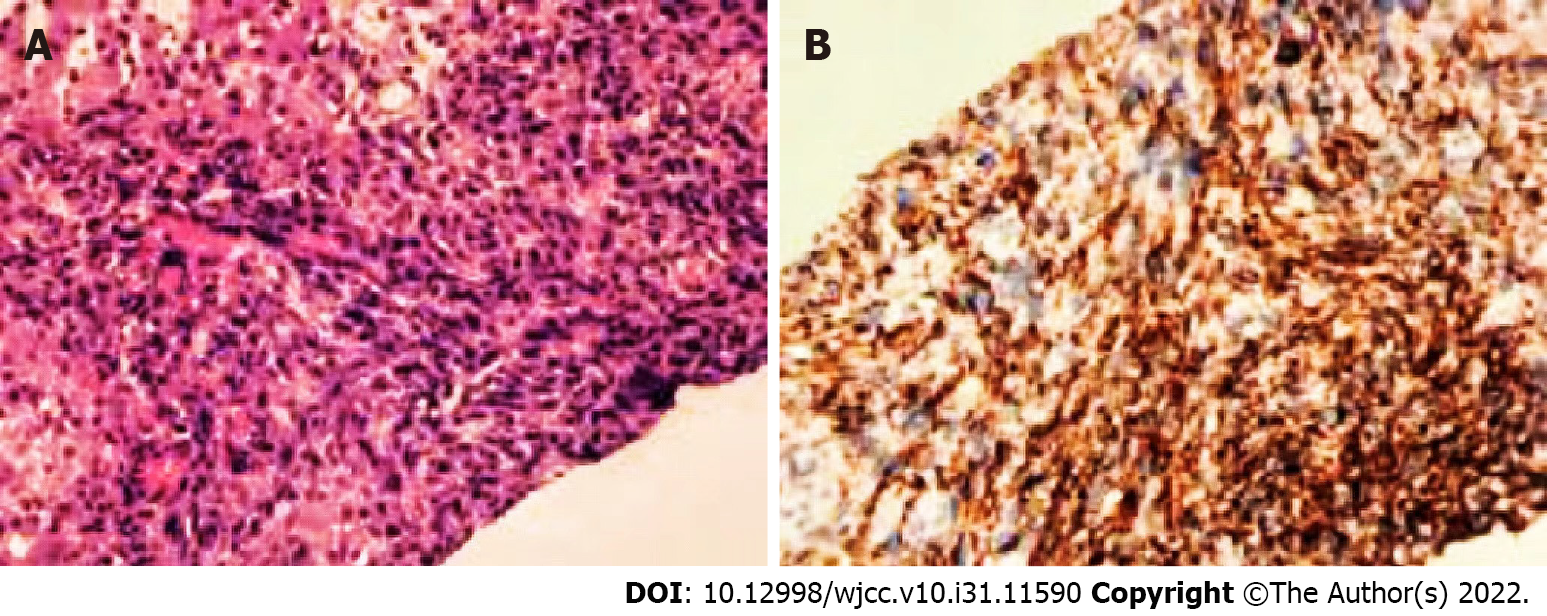
Figure 4 Histopathology reveals angiosarcoma of vascular origin.
A: Obvious pleomorphic and heterotypic tumor cells arranged in clusters (magnification 100 ×); B: Immunohistochemical staining (magnification 100 ×) reveals CD31 positivity.
- Citation: Wang J, Sun LT. Primary hepatic angiosarcoma: A case report. World J Clin Cases 2022; 10(31): 11590-11596
- URL: https://www.wjgnet.com/2307-8960/full/v10/i31/11590.htm
- DOI: https://dx.doi.org/10.12998/wjcc.v10.i31.11590


