Copyright
©The Author(s) 2022.
World J Clin Cases. Jan 21, 2022; 10(3): 992-999
Published online Jan 21, 2022. doi: 10.12998/wjcc.v10.i3.992
Published online Jan 21, 2022. doi: 10.12998/wjcc.v10.i3.992
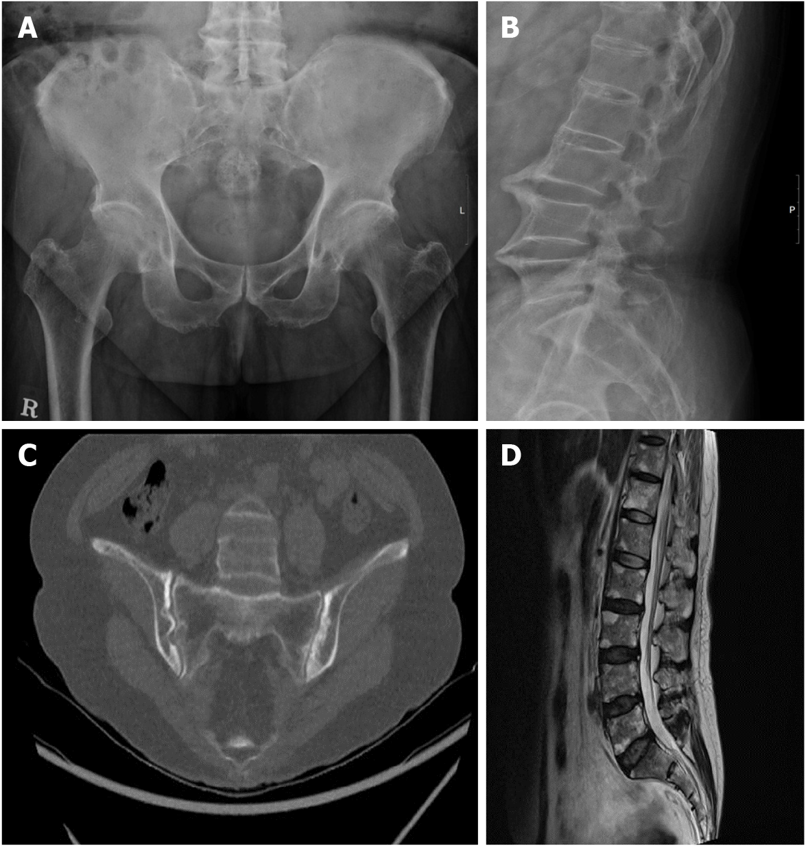
Figure 1 Radiographic images of the father (case 1) and the daughter (case 2) with ankylosing spondylitis.
A: Sacroiliac joint of the father showing complete ankyloses; B: Lumbar spine imaging of the father in the shape of “bamboo spine”; C: Computed tomography image of the sacroiliac joint of the daughter showing multiple definite erosions with sclerotic changes on both sacroiliac joints; D: Magnetic resonance image of the lumbar spine of the daughter revealing fat deposition at the corners of the lumbar spine, probably due to ankylosing spondylitis.
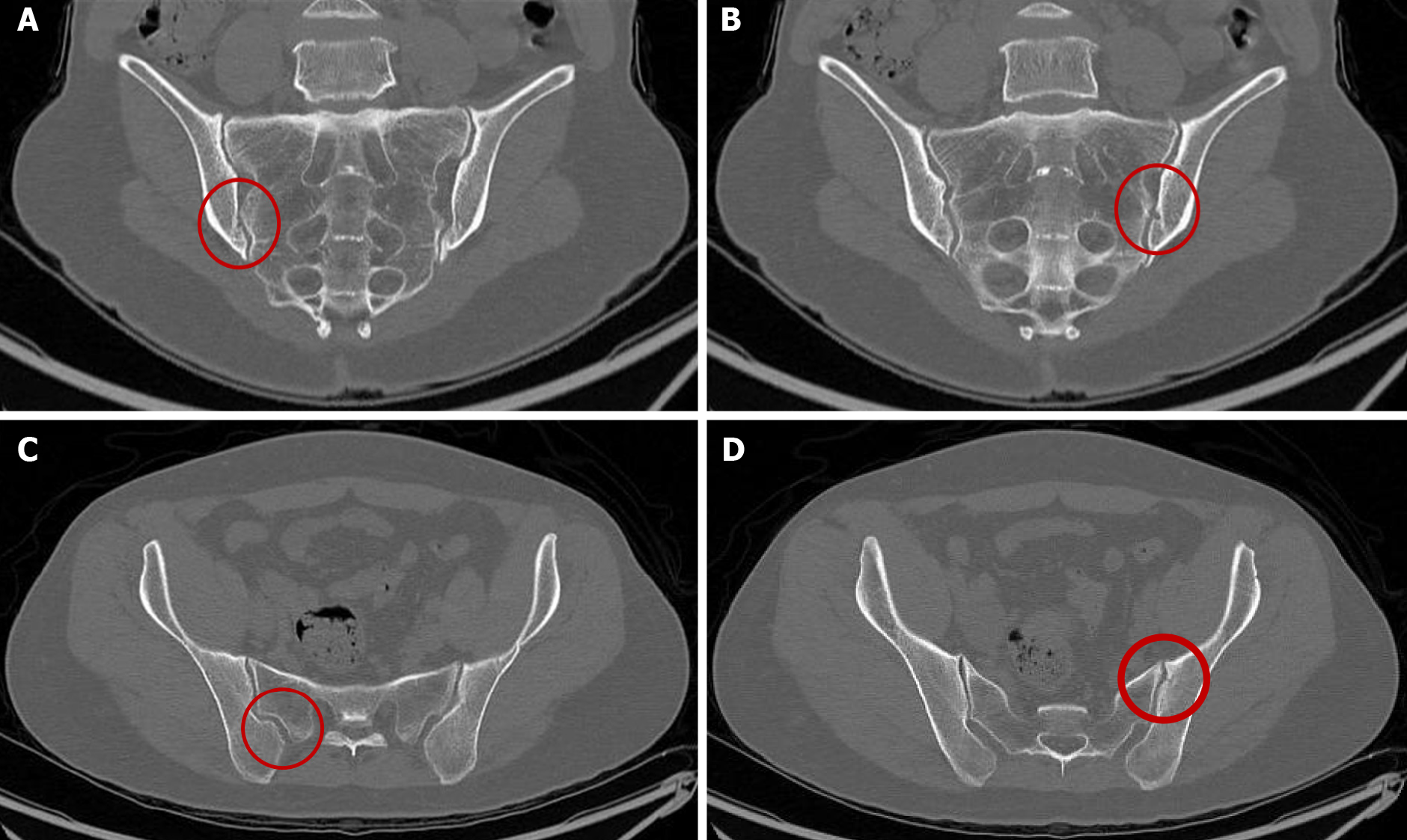
Figure 2 Computed tomography image of the sacroiliac joint(s) of the two daughters who tested positive for human leukocyte antigen B51.
A, B: Multiple small undulating lesions are apparent on both sacroiliac joints, suggesting grade 1 bilateral sacroiliitis were seen in one daughter; C, D: Small undulating lesions in both sacroiliac joints are apparent, suggesting grade 1 bilateral sacroiliitis in the other daughter. The lesions are indicated by the red circle.
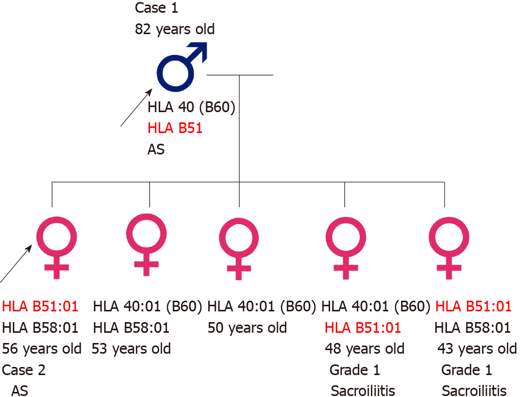
Figure 3 Pedigree of the family.
Human leukocyte antigen B51:01 carriers had either ankylosing spondylitis (black arrow) or grade 1 sacroiliitis. HLA: Human leukocyte antigen.
- Citation: Lim MJ, Noh E, Lee RW, Jung KH, Park W. Occurrence of human leukocyte antigen B51-related ankylosing spondylitis in a family: Two case reports. World J Clin Cases 2022; 10(3): 992-999
- URL: https://www.wjgnet.com/2307-8960/full/v10/i3/992.htm
- DOI: https://dx.doi.org/10.12998/wjcc.v10.i3.992


