Copyright
©The Author(s) 2022.
World J Clin Cases. Jan 21, 2022; 10(3): 1077-1085
Published online Jan 21, 2022. doi: 10.12998/wjcc.v10.i3.1077
Published online Jan 21, 2022. doi: 10.12998/wjcc.v10.i3.1077
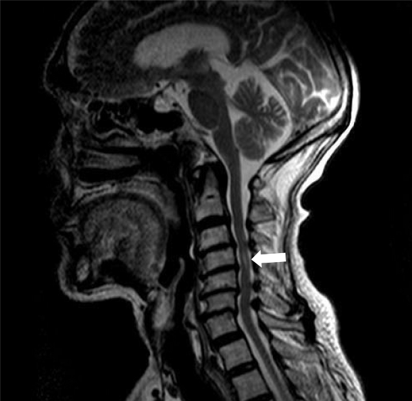
Figure 1 Magnetic resonance imaging of the cervical spine.
Cervical syringomyelia at C4 (white arrow); cervical disc herniation and spinal stenosis from the C3 to C7 Levels.
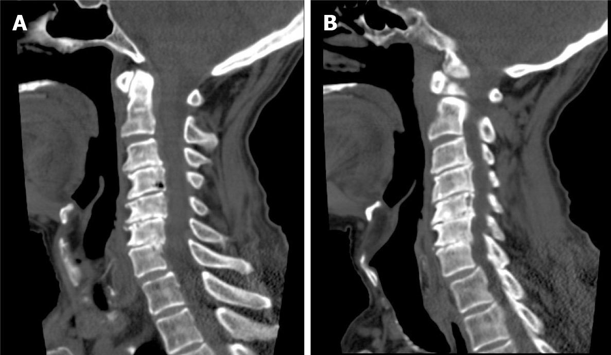
Figure 2 Computed tomography of the cervical spine.
A: C4 vertebral body destruction; B: C5-7 vertebral body assimilation.
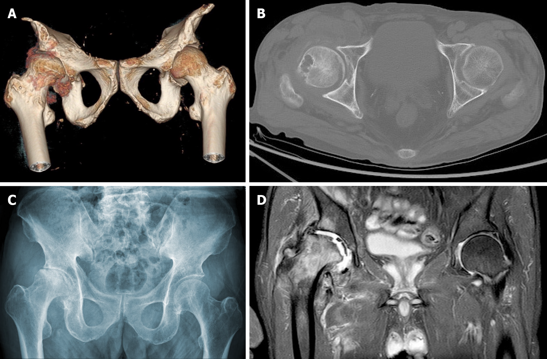
Figure 3 Right hip joint.
A: Computed tomography (CT) three-dimensional reconstruction; B: CT; C: X-ray. Joint space loss, articular surface collapse, and destructive acetabulum and femoral head changes; D: Right hip magnetic resonance imaging, T2W1. Articular cartilage loss, joint degeneration, soft tissue disorder, and obvious joint fluid were observed.
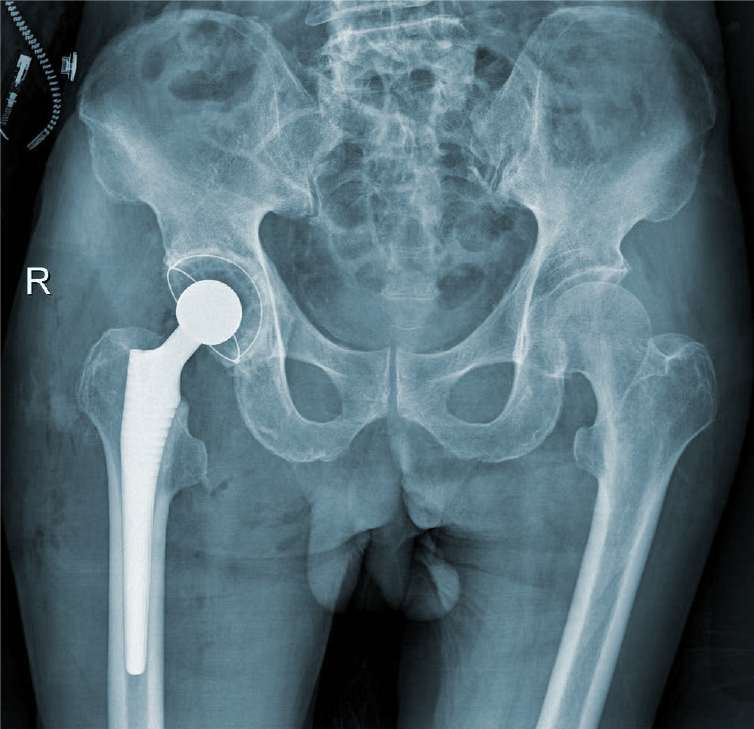
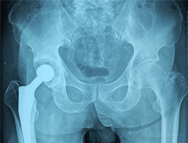
Figure 4 Right hip X-ray film after total hip arthroplasty.
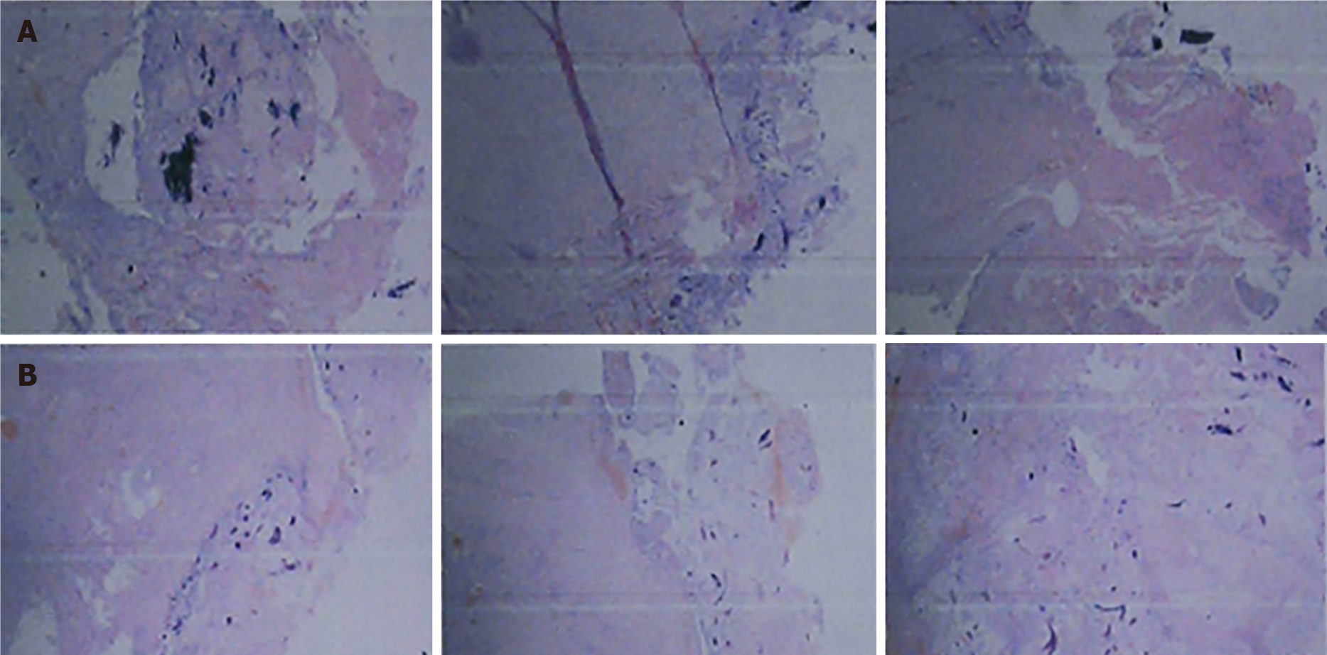
Figure 5 Pathological results.
A: Right femoral head tissue sections during surgery. Tissue degeneration, calcification, small vascular proliferation and a multicore giant cell response in some regions; B: Synovial tissue sections of the right hip joint during the operation. Capillary hyperplasia was accompanied by infiltration of acute chronic inflammatory cells, local region tissue denaturation and calcification.
Figure 6 Follow-up right hip X-ray film 6 mo after total hip arthroplasty.
X-rays revealed that the acetabular inclination and anteversion were well maintained and showed signs of periprosthetic bone growth without clinical manifestations of implant loosening compared with postoperative observations.
- Citation: Lu Y, Xiang JY, Shi CY, Li JB, Gu HC, Liu C, Ye GY. Cervical spondylotic myelopathy with syringomyelia presenting as hip Charcot neuroarthropathy: A case report and review of literature. World J Clin Cases 2022; 10(3): 1077-1085
- URL: https://www.wjgnet.com/2307-8960/full/v10/i3/1077.htm
- DOI: https://dx.doi.org/10.12998/wjcc.v10.i3.1077


