Copyright
©The Author(s) 2022.
World J Clin Cases. Jan 21, 2022; 10(3): 1032-1040
Published online Jan 21, 2022. doi: 10.12998/wjcc.v10.i3.1032
Published online Jan 21, 2022. doi: 10.12998/wjcc.v10.i3.1032
Figure 1 Thyroid ultrasonography.
A: Solid nodule with multiple punctate microcalcifications and relatively regular shape within the right lobe of the thyroid (white arrow); B: A huge cystic mass with a clear boundary located in the lower right lobe of the thyroid (white arrow).
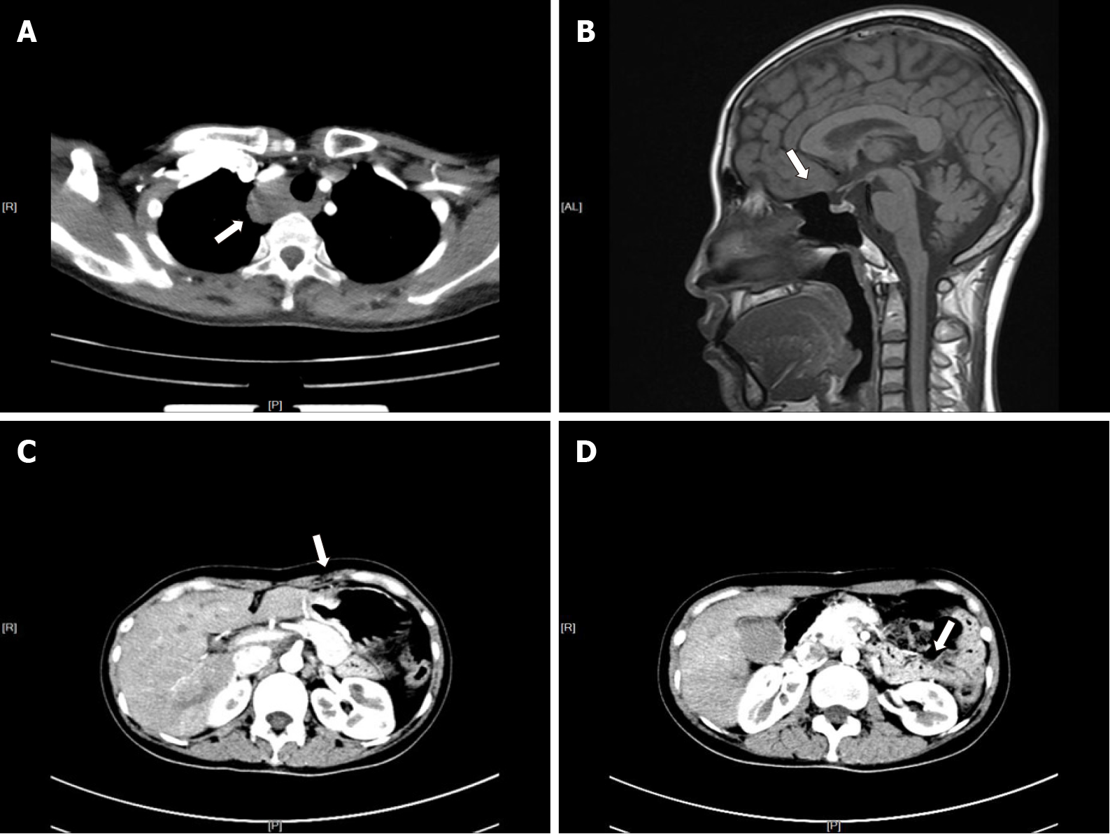
Figure 2 Computed tomography/magnetic resonance imaging examination.
A: A lesion located in the right side of the trachea (white arrow); B: Enlarged pituitary structure (white arrow); C: Remnant stomach anastomosed to the jejunum (white arrow); D: Remnant pancreas body and tail (white arrow).
Figure 3 Single-photon emission computed tomography/computed tomography and intraoperative findings.
A: Single-photon emission computed tomography/computed tomography revealed that a mass was located below the lower pole of the right thyroid lobe that was growing downward and protruding into the superior mediastinum. No significant radioactivity concentration was observed in the mass (white arrow); B: A large cyst was filled with clear watery fluid and contiguous with the lower right thyroid lobe (white arrow).
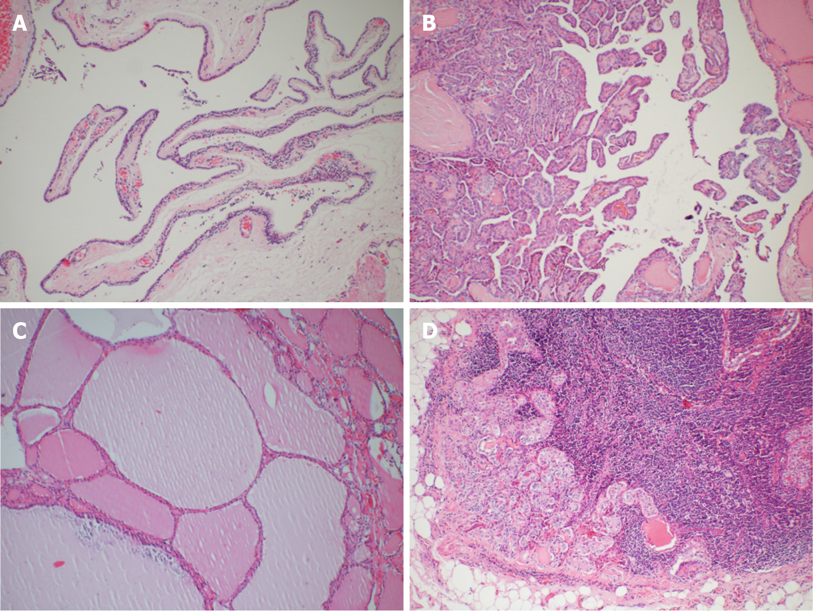
Figure 4 Histopathological examinations.
A: Parathyroid cyst: a single locular cystic mass covered by a single layer of flattened transparent cells with small clusters of extruded parathyroid tissue in the wall; B: Thyroid papillary carcinoma: complex branching papilla with fibrous vascular center, and surface coated with simple columnar epithelium. The epithelial nuclei were ground-glass like, with nuclear grooves, intranuclear pseudo-inclusions, and nuclear overlap; C: Nodular goiter: the follicles vary in size and are filled with colloid; D: Metastatic lesions of thyroid papillary carcinoma in lymph nodes. Hematoxylin and eosin staining, 100× magnification.
- Citation: Xu JL, Dong S, Sun LL, Zhu JX, Liu J. Multiple endocrine neoplasia type 1 combined with thyroid neoplasm: A case report and review of literatures. World J Clin Cases 2022; 10(3): 1032-1040
- URL: https://www.wjgnet.com/2307-8960/full/v10/i3/1032.htm
- DOI: https://dx.doi.org/10.12998/wjcc.v10.i3.1032


