Copyright
©The Author(s) 2022.
World J Clin Cases. Jan 21, 2022; 10(3): 1024-1031
Published online Jan 21, 2022. doi: 10.12998/wjcc.v10.i3.1024
Published online Jan 21, 2022. doi: 10.12998/wjcc.v10.i3.1024
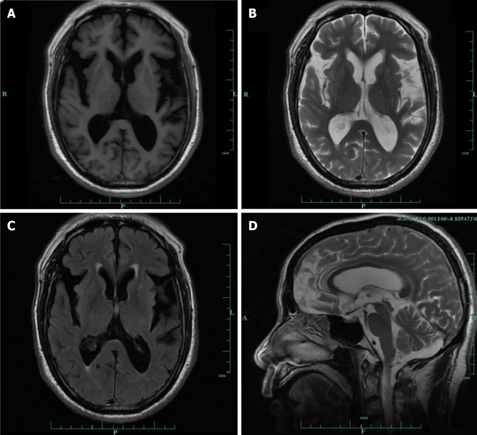
Figure 1 Magnetic resonance imaging shows mild bilateral frontotemporal atrophy.
A-C: The T1, T2 and FLAIR sequence showed temporal atrophy, with broadening of the posterior horn of the lateral ventricle; D: Magnetic resonance sagittal view showed mild frontal lobe atrophy.
- Citation: Xu T, Li ZS, Fang W, Cao LX, Zhao GH. Concomitant Othello syndrome and impulse control disorders in a patient with Parkinson’s disease: A case report. World J Clin Cases 2022; 10(3): 1024-1031
- URL: https://www.wjgnet.com/2307-8960/full/v10/i3/1024.htm
- DOI: https://dx.doi.org/10.12998/wjcc.v10.i3.1024


