Copyright
©The Author(s) 2022.
World J Clin Cases. Oct 6, 2022; 10(28): 9985-10003
Published online Oct 6, 2022. doi: 10.12998/wjcc.v10.i28.9985
Published online Oct 6, 2022. doi: 10.12998/wjcc.v10.i28.9985
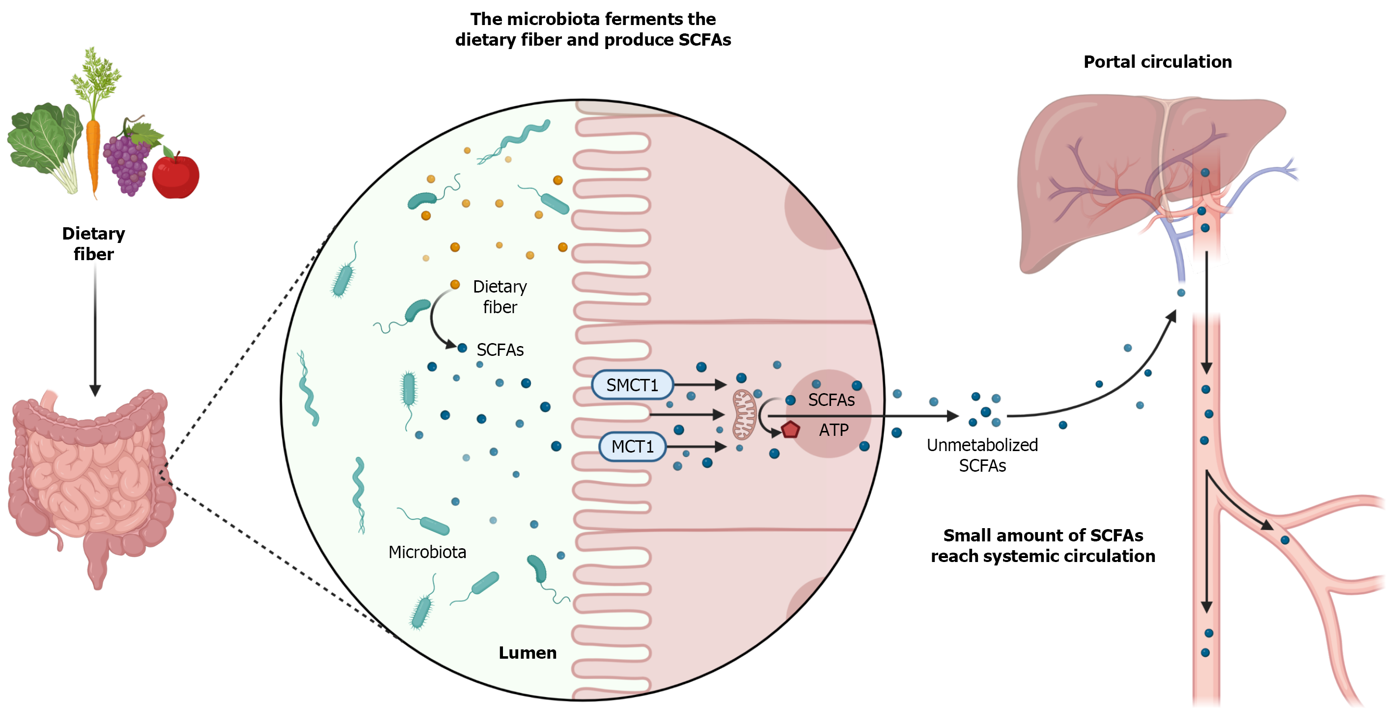
Figure 1 Production and metabolism of shortchain fatty acids.
Short-chain fatty acids (SCFAs) are produced by the intestinal microbiota from the fermentation of dietary fibers. SCFAs are absorbed by colonocytes mainly through monocarboxylate transporter 1 (MCT1), sodium-dependent monocarboxylate transporter 1 (SMCT1) and passive diffusion. At intracellular levels, SCFAs are metabolized and converted into ATP that will serve as energy source for these cells. Unmetabolized SCFAs cross the basolateral membrane and reach the portal circulation. In the liver, SCFAs are used in the synthesis of cholesterol and glucose. Only a small amount of SCFAs not used by the liver reach the systemic circulation[221]. The authors have obtained the permission for figure using from the BioRender.com (Supplementary material).
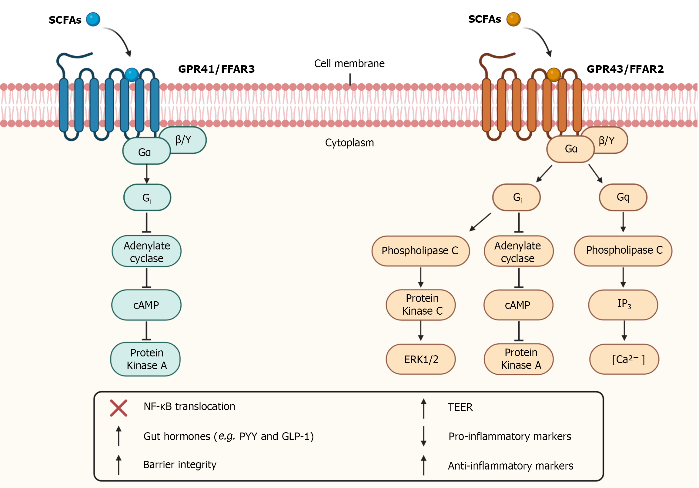
Figure 2 Short-chain fatty receptors and intracellular signaling.
Short-chain fatty acids (SCFAs) binds to G-protein coupled receptors such as GPR41 and GPR43 receptors also known as free fatty acid receptors (FFAR) 3 and FFAR2, respectively. The activation of GPR41 and GPR43 receptors through Gi subunit promotes inhibition of adenylate cyclase which inhibit cyclic adenosine monophosphate (cAMP) that inhibit protein kinase A. GPR43 receptor activation through Gq subunit promotes activation of phospholipase C which activate inositol 1,4,5-trisphosphate (IP3) leading to the elevation of intracellular calcium levels. GPR43 receptor activation through Gq subunit also promotes activation of phospholipase C which activate protein kinase C leading to activation of extracellular signal-regulated kinase 1/2 (ERK1/2) cascade. The activation of these receptors inhibits translocation of nuclear factor kappa B (NF- ΚB), altering the expression of certain proteins, promotes an increase in the release of gut hormones such as Peptide YY (PYY) and Glucagon-like peptide 1 (GLP-1), increase intestinal barrier integrity and Transepithelial Electrical Resistance (TEER), reduce pro-inflammatory markers levels, and increase anti-inflammatory markers levels. The authors have obtained the permission for figure using from the BioRender.com (Supplementary material).
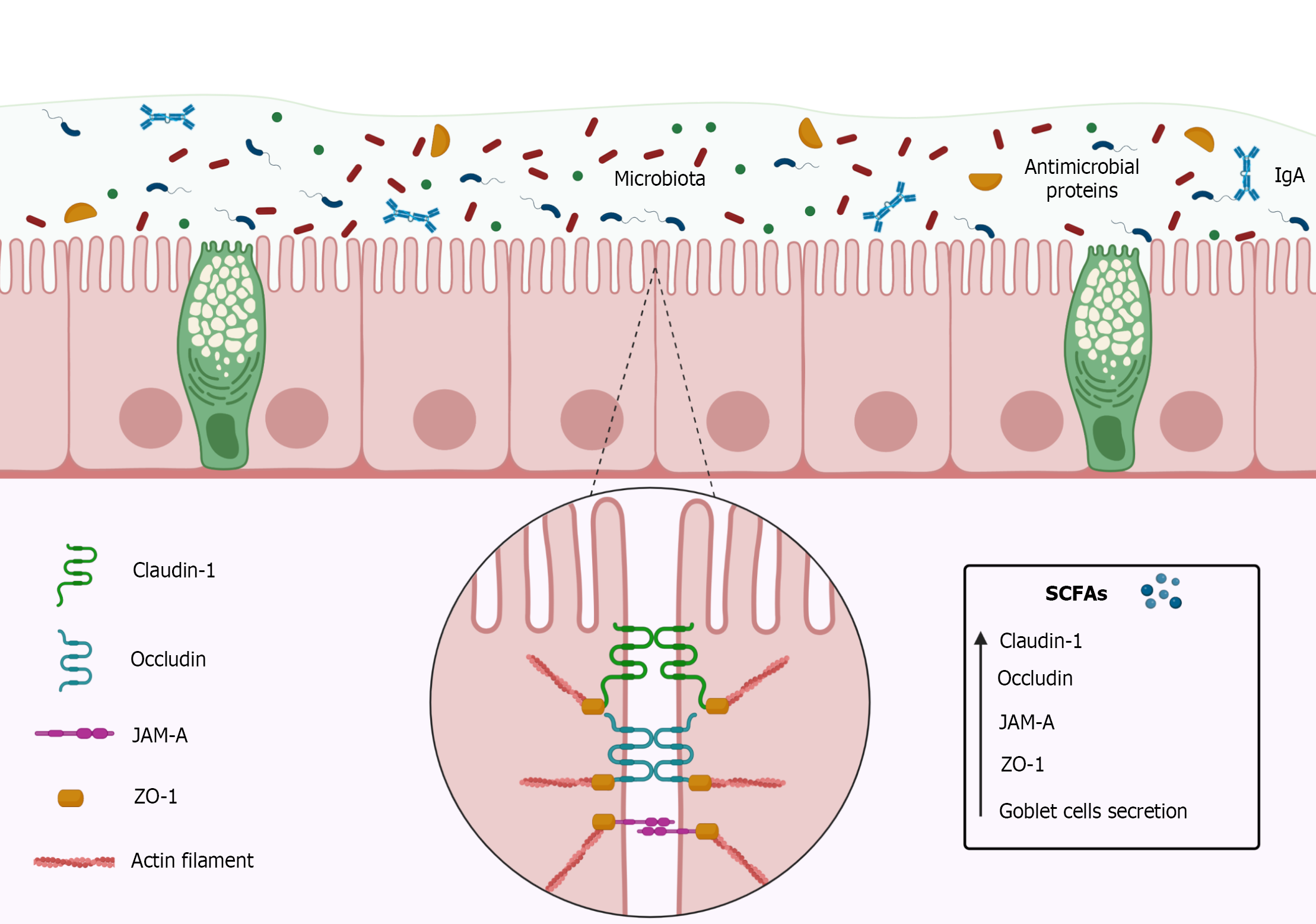
Figure 3 Intestinal barrier constitution and short-chain fatty acids effects.
The intestinal barrier is composed by simple cylindrical epithelial cells, mucus, microbiota, immunoglobulins A (IgA), antimicrobial proteins, and intercellular binding proteins such as tight junction proteins. Tight junctions play an important role in the integrity of the intestinal barrier by keeping epithelial cells well adhered to each other, and are composed of transmembrane proteins such as Claudins, Occludin, Junctional adhesion molecules (JAM), and accessory cytoplasmic proteins such as Zonulas Occludens (ZOs). Claudin-1 pairs with other Claudin-1 Loops of the adjacent cell, decreasing paracellular permeability. Like Claudin-1, Occludin pairs with other Occludin loops of the adjacent cell forming a barrier mainly to macromolecules. JAM-A belongs to immunoglobulins superfamily and can form homophilic interactions adjacent to tight junctions and interacting with integrins or other members of the JAM family. ZO-1 are located on the cytoplasm and perform the anchorage of proteins joining them to the cytoskeleton through actin filaments. Short-chain fatty acids (SCFAs), mainly Butyrate, are capable of increase Claudin-1, Occludin, JAM, ZO proteins and increase mucus secretion by goblet cells. The authors have obtained the permission for figure using from the BioRender.com (Supplementary material).
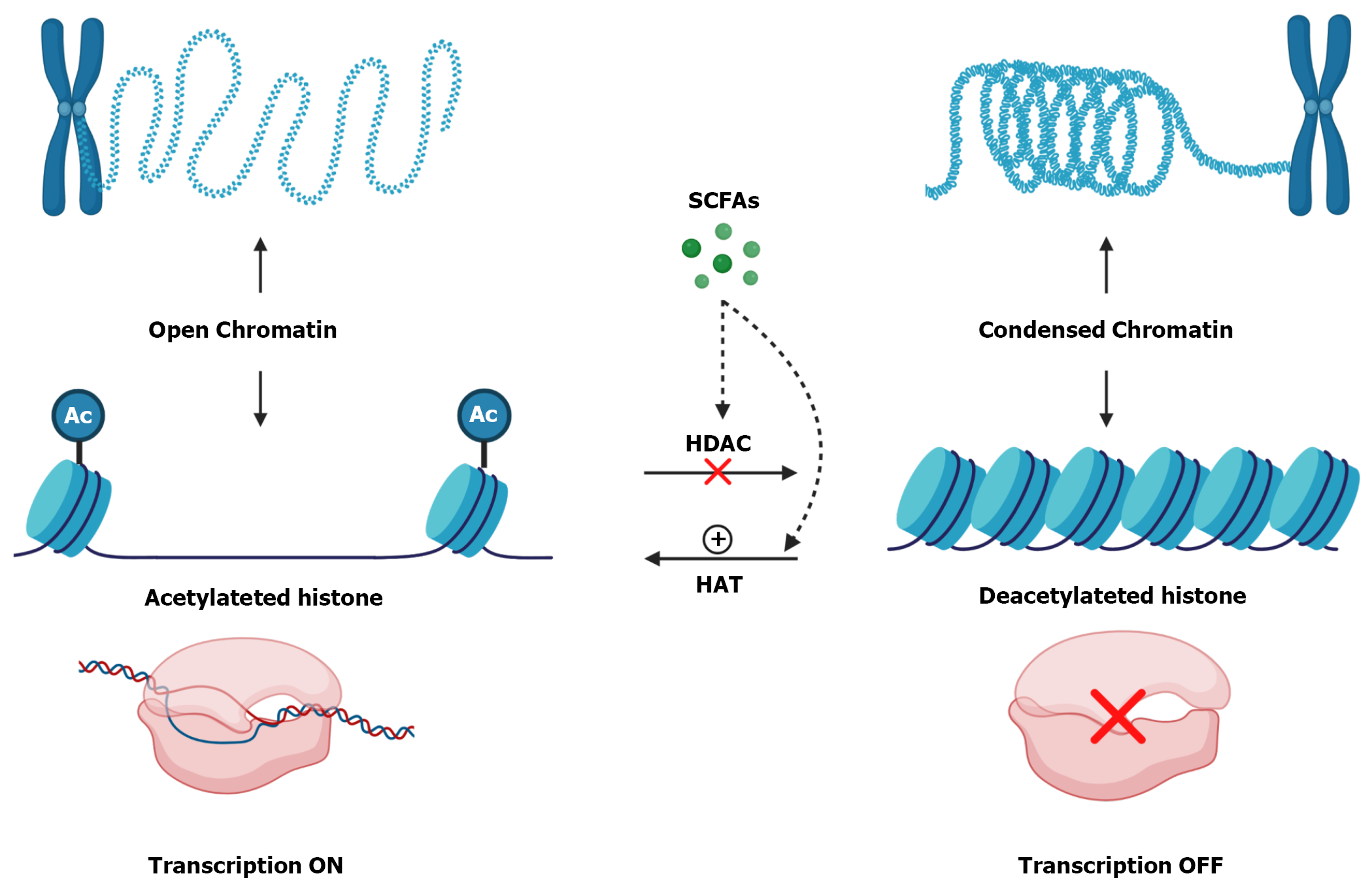
Figure 4 Histone modifications and short-chain fatty acids influence.
Histones are proteins that interacts with DNA and play an important role in organizing the double strand. When histones contain acetyl (Ac) groups they become acetylated and there is a repulsion between these groups, so histone becomes more distant from each other, causing chromatin to become decondensed and, consequently, more accessible which activate transcription. Otherwise, when histones are deacetylated, without Ac groups, DNA becomes more tangled which makes chromatin more condensed and promotes gene silencing. Histone Deacetylases (HDAC) are enzymes that remove Ac groups from Histones making them deacetylated. Otherwise, Histone Acetylases (HAT) are enzymes that insert Ac groups in Histones making them acetylated. Short-chain fatty acids (SCFAs), mainly Butyrate, inhibit HDAC and increase HAT activity. The authors have obtained the permission for figure using from the BioRender.com (Supplementary material).
- Citation: Caetano MAF, Castelucci P. Role of short chain fatty acids in gut health and possible therapeutic approaches in inflammatory bowel diseases. World J Clin Cases 2022; 10(28): 9985-10003
- URL: https://www.wjgnet.com/2307-8960/full/v10/i28/9985.htm
- DOI: https://dx.doi.org/10.12998/wjcc.v10.i28.9985


