Copyright
©The Author(s) 2022.
World J Clin Cases. Oct 6, 2022; 10(28): 10375-10383
Published online Oct 6, 2022. doi: 10.12998/wjcc.v10.i28.10375
Published online Oct 6, 2022. doi: 10.12998/wjcc.v10.i28.10375
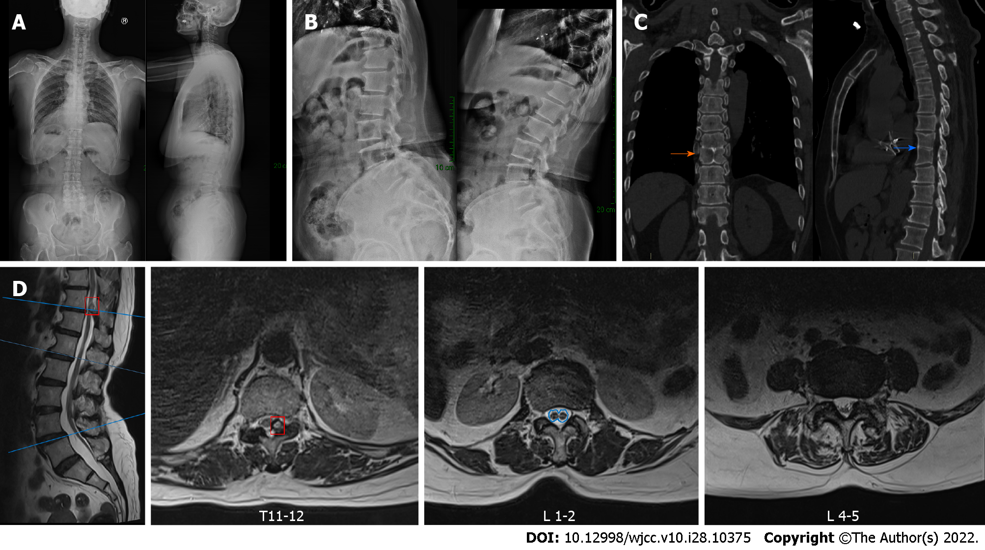
Figure 1 Preoperative radiography.
A: Preoperative anteroposterior and lateral radiographs; B: Preoperative flexion–extension radiographs; C: Preoperative computed tomography scan showing block vertebrae at T8/T9 (orange arrow), butterfly vertebra atT9 (blue arrow); D: Preoperative magnetic resonance imaging showing thoracic ossification of the ligamentum flavum (red box), a split cord malformation (blue outline) with a low-lying conus and a thickened terminal filament deposited with fatty tissue, and bilateral nerve root compression at L4-5.
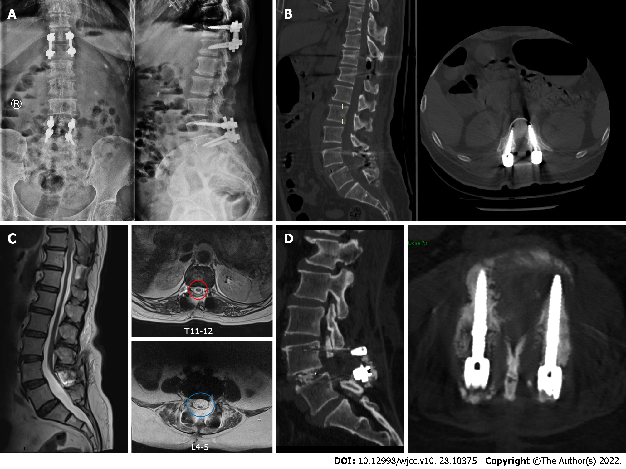
Figure 2 Radiography after surgery and at follow-up.
A: Postoperative anteroposterior and lateral thoracolumbar radiographs; B: Postoperative thoracolumbar computed tomography (CT) scan showing the absence of thoracic ossification of the ligamentum flavum; C: Magnetic resonance imaging at 1 mo after surgery showing good decompression of the spinal canal (red and blue circle); D: Lumbar CT scan at 3 mo after surgery showing the intervertebral fusion status and good position of screws.
- Citation: Wang YT, Mu GZ, Sun HL. Thoracolumbar surgery for degenerative spine diseases complicated with tethered cord syndrome: A case report. World J Clin Cases 2022; 10(28): 10375-10383
- URL: https://www.wjgnet.com/2307-8960/full/v10/i28/10375.htm
- DOI: https://dx.doi.org/10.12998/wjcc.v10.i28.10375


