Copyright
©The Author(s) 2022.
World J Clin Cases. Oct 6, 2022; 10(28): 10358-10365
Published online Oct 6, 2022. doi: 10.12998/wjcc.v10.i28.10358
Published online Oct 6, 2022. doi: 10.12998/wjcc.v10.i28.10358
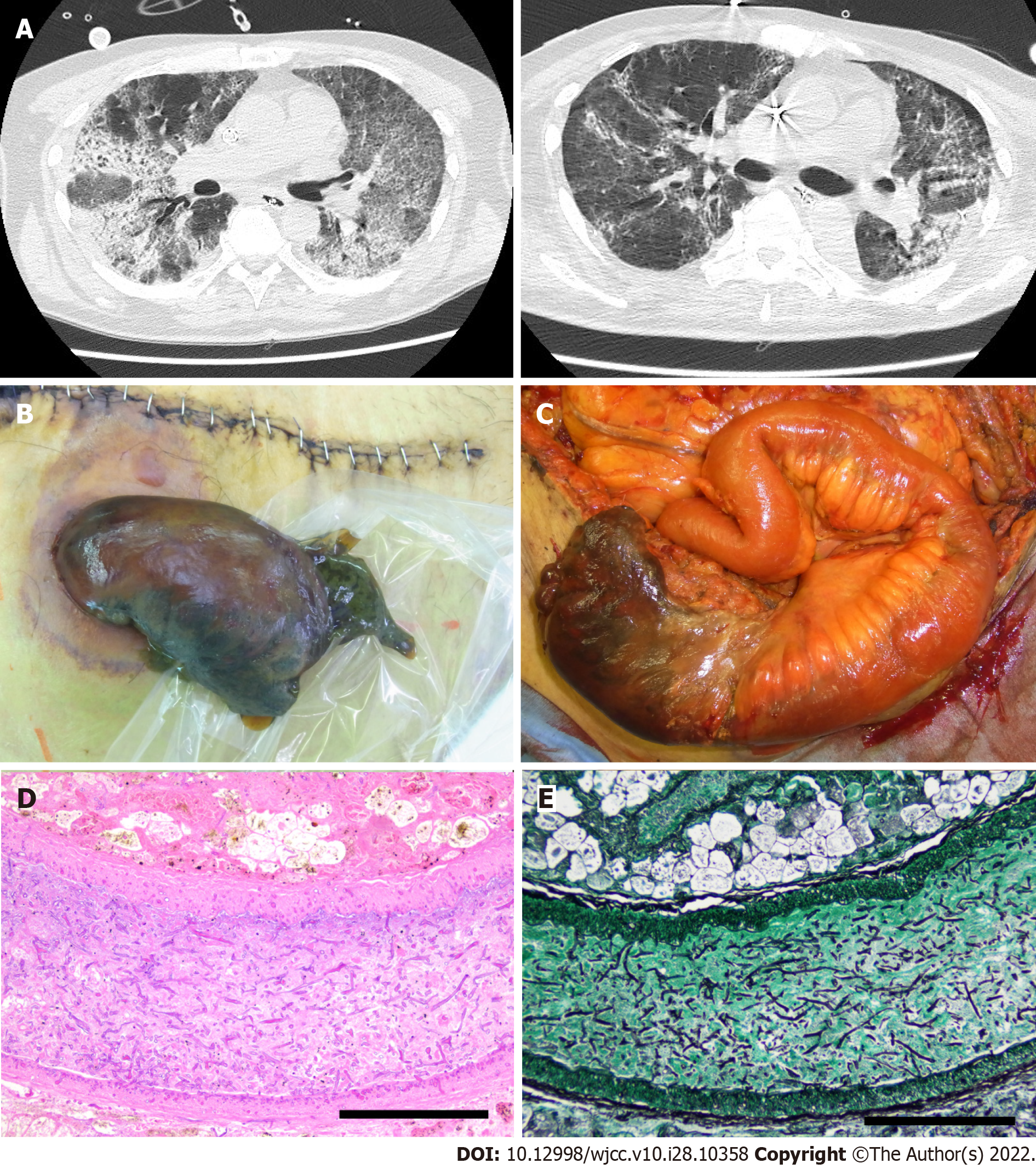
Figure 1 Imaging and histological findings during treatment.
A: Computed tomography (CT) images of acute respiratory distress syndrome (ARDS). Left image: CT image at the time of ARDS diagnosis. Right image: CT image after 38 d; B: The necrotic ileostoma caused by the mucormycosis can be seen in this image; C: Surgical findings at the stomal reconstruction; D and E; Presence of thrombus with Mucorales in the mesenteric vessels of the necrotic ileostoma. H&E staining; magnification × 200 (D), Grocott staining; magnification × 200 (E). Bar: 200 μm.
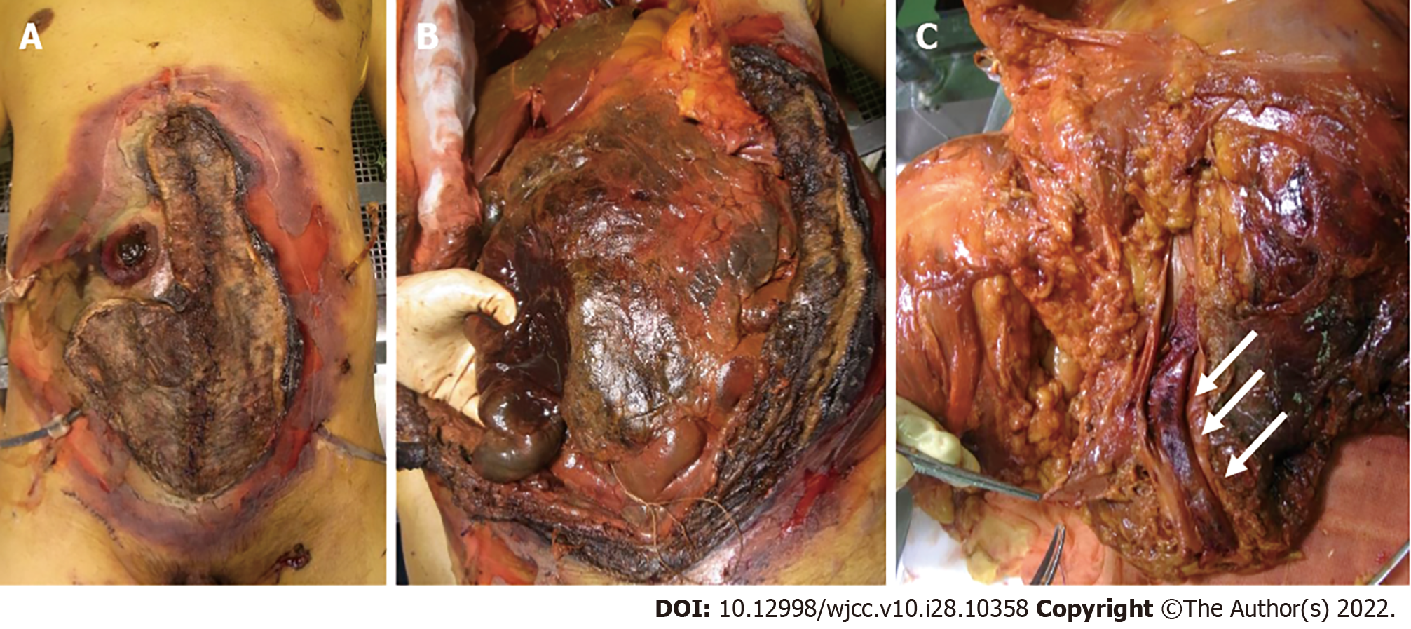
Figure 2 Autopsy findings of the Mucorales infection.
A: The image shows the necrotic stoma, skin, and abdominal wall; B: The necrotic abdominal organs are seen in this image. A pathologist held the intestine on the oral side of the stoma; C: Dorsal view of the incised common iliac vein. The thrombus can be seen in the common iliac vein (indicated by arrows).
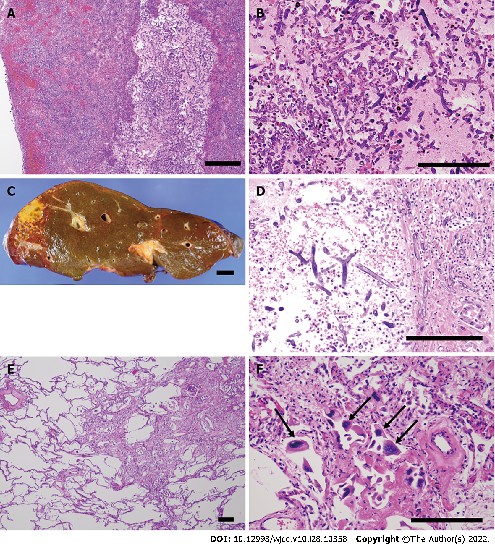
Figure 3 Histopathological findings of the Mucorales infection in the organs and thrombus.
A and B: Presence of Mucorales in the thrombus; H&E staining; magnification × 100 (A), × 200 (B); C: Macroscopic image showing partial necrosis of the liver; D: Hepatic infarction caused by the thrombus including Mucorales; H&E staining; magnification × 200; E: The proliferative/organizing phase of diffuse alveolar damage of the left lung. Restoration of type II pneumocytes and proliferation of myofibroblasts are partially shown. H&E staining; magnification × 40; F: Cytomegalovirus infection. Cytomegalovirus-infected cells are indicated by black arrows. H&E staining; magnification × 200. Bar in the autopsy images: 2 cm; Bar in the microscopic images: 200 μm.
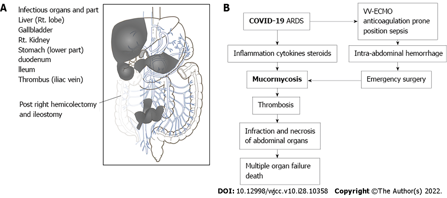
Figure 4 The autopsy diagnosis.
A: The image shows the organs and part of the Mucorales infection. The gray areas indicate areas infected with Mucorales. The resected ascending colon is transparent in the drawing; B: A flowchart of the autopsy diagnosis.
- Citation: Kyuno D, Kubo T, Tsujiwaki M, Sugita S, Hosaka M, Ito H, Harada K, Takasawa A, Kubota Y, Takasawa K, Ono Y, Magara K, Narimatsu E, Hasegawa T, Osanai M. COVID-19-associated disseminated mucormycosis: An autopsy case report. World J Clin Cases 2022; 10(28): 10358-10365
- URL: https://www.wjgnet.com/2307-8960/full/v10/i28/10358.htm
- DOI: https://dx.doi.org/10.12998/wjcc.v10.i28.10358


