Copyright
©The Author(s) 2022.
World J Clin Cases. Oct 6, 2022; 10(28): 10339-10345
Published online Oct 6, 2022. doi: 10.12998/wjcc.v10.i28.10339
Published online Oct 6, 2022. doi: 10.12998/wjcc.v10.i28.10339
Figure 1 Chromosome karyotyping analysis.
A and B: G-banded karyotyping of bone marrow mononucleated cells upon detection of anaplastic large-cell lymphoma transformation revealed complex cytogenetic abnormalities in t(9;22)(q34;q11) as follows: 49-51, XX, +1, -7, +8, t(9; 22)(q34; q11), +10, +11, add(16)(p13), ? add(17)(p11), +18, +21, -22, +mar, 1min, inc [cp10]/46, XX[10].
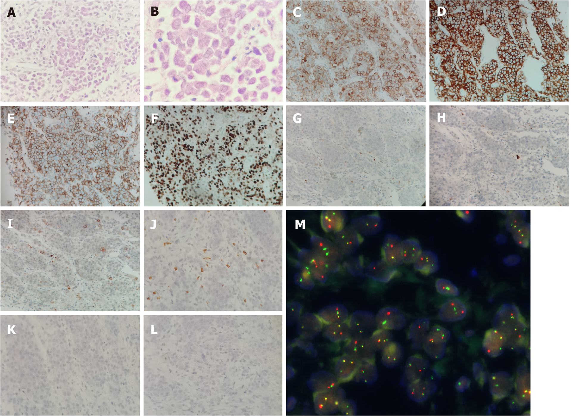
Figure 2 Histological and immunohistochemical analysis of the mass.
A and B: Haematoxylin and eosin (HE) staining showed diffused medium-sized malignant cells with irregular nuclear contours, vesicular nuclei, and prominent nucleoli (A: × 40, B: × 100); C-F: The malignant cells were positive for CD30, CD4, CD43, and Ki67 respectively (× 40); G-L: The malignant cells were negative for anaplastic lymphoma kinase, myeloperoxidase, CD20, CD3, T-cell-restricted intracellular antigen, and CD235a respectively (× 40); M: Fluorescence in situ hybridization (FISH) of a paraffin-embedded biopsy specimen revealed the presence of BCR and ABL rearrangements in the malignant lymphoma cells. Red signals represent the ABL gene, green signals the BCR gene, and yellow signals the BCR-ABL fusion gene (× 1000).
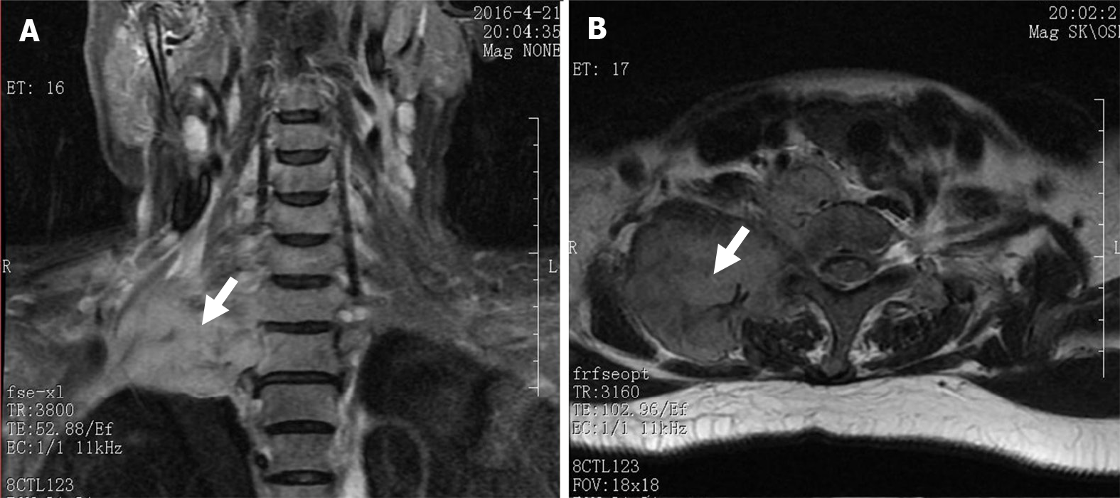
Figure 3 Magnetic resonance image of the shoulder.
A large soft tissue mass (white arrows) is evident in the right cervical root and supraclavicular fossa. A: Coronal plane; B: Transverse plane.
- Citation: Wu Q, Kang Y, Xu J, Ye WC, Li ZJ, He WF, Song Y, Wang QM, Tang AP, Zhou T. Sudden extramedullary and extranodal Philadelphia-positive anaplastic large-cell lymphoma transformation during imatinib treatment for CML: A case report. World J Clin Cases 2022; 10(28): 10339-10345
- URL: https://www.wjgnet.com/2307-8960/full/v10/i28/10339.htm
- DOI: https://dx.doi.org/10.12998/wjcc.v10.i28.10339


