Copyright
©The Author(s) 2022.
World J Clin Cases. Oct 6, 2022; 10(28): 10326-10331
Published online Oct 6, 2022. doi: 10.12998/wjcc.v10.i28.10326
Published online Oct 6, 2022. doi: 10.12998/wjcc.v10.i28.10326
Figure 1 Axial and coronal noncontrast computed tomography scans with soft windows showing an opacified left posterior ethmoidal cell with a thickened bony shell.
Across the lamina papyracea was a lesion with a fusiform change and isodensity. A: Axial plane; B: Coronal plane.
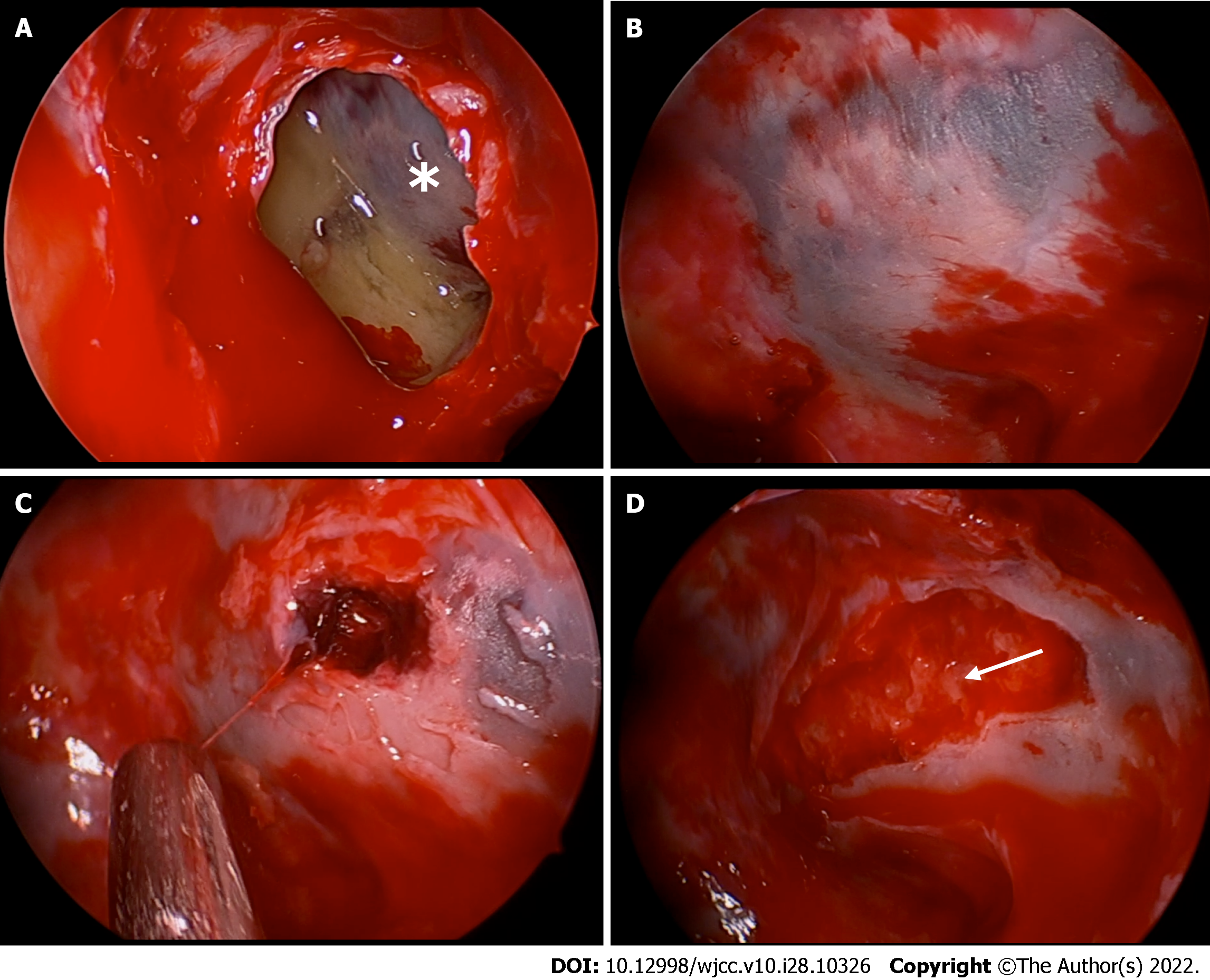
Figure 2 Endoscopic views of the mucocele and the subperiosteal orbital collection.
A: Marsupialization of the mucocele of the left posterior ethmoid sinus. An asterisk indicates the lamina papyracea; B: The lamina papyracea is covered with thin devitalized mucosa and has a blue and white appearance with a geographic pattern. The surrounding mucosa rapidly became hyperemic after the cavity pressure was eliminated; C: A blood clot was uncovered under the blue area of the lamina papyracea, and the periorbita was subsequently exposed; D: Pus (white arrow) oozed from around the blood clot.
Figure 3 Postoperative magnetic resonance imaging and follow-up with endoscopy.
A: T2 magnetic resonance imaging scan on the 5th postoperative day shows hyperintensity of the involved medial periorbita of the left orbit on T2-weighted imaging, revealing sufficient drainage of the subperiosteal orbital abscess; B: Endoscopic examination on the 11th postoperative day shows a slightly bulging periorbita that was apparently healthy; C: Endoscopic reevaluation two months after the operation revealed nearly complete epithelization of the lamina papyracea with a small cherry granuloma.
- Citation: Hu XH, Zhang C, Dong YK, Cong TC. Subperiosteal orbital hematoma concomitant with abscess in a patient with sinusitis: A case report. World J Clin Cases 2022; 10(28): 10326-10331
- URL: https://www.wjgnet.com/2307-8960/full/v10/i28/10326.htm
- DOI: https://dx.doi.org/10.12998/wjcc.v10.i28.10326


