Copyright
©The Author(s) 2022.
World J Clin Cases. Oct 6, 2022; 10(28): 10293-10300
Published online Oct 6, 2022. doi: 10.12998/wjcc.v10.i28.10293
Published online Oct 6, 2022. doi: 10.12998/wjcc.v10.i28.10293
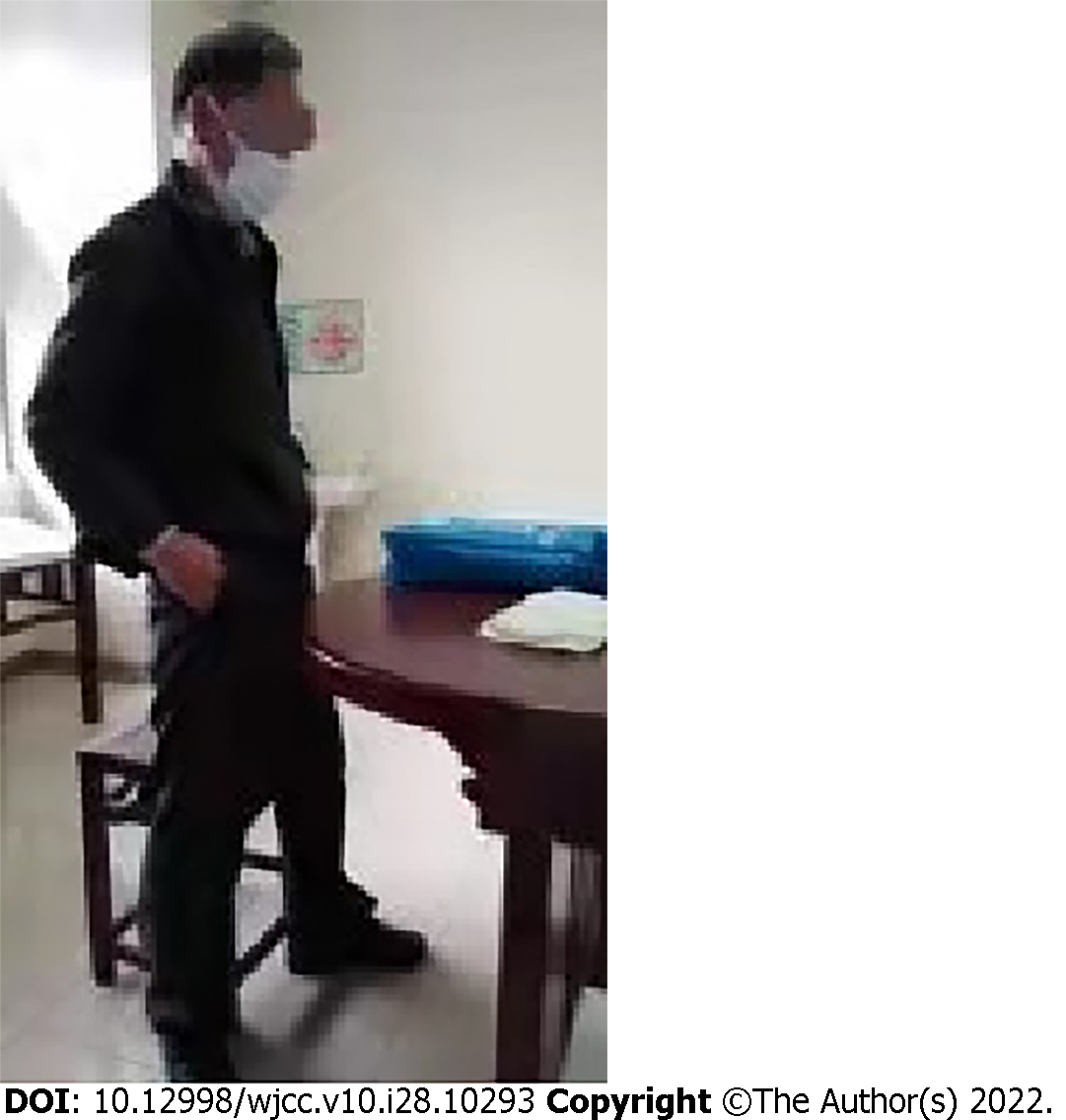
Figure 1
Body size.
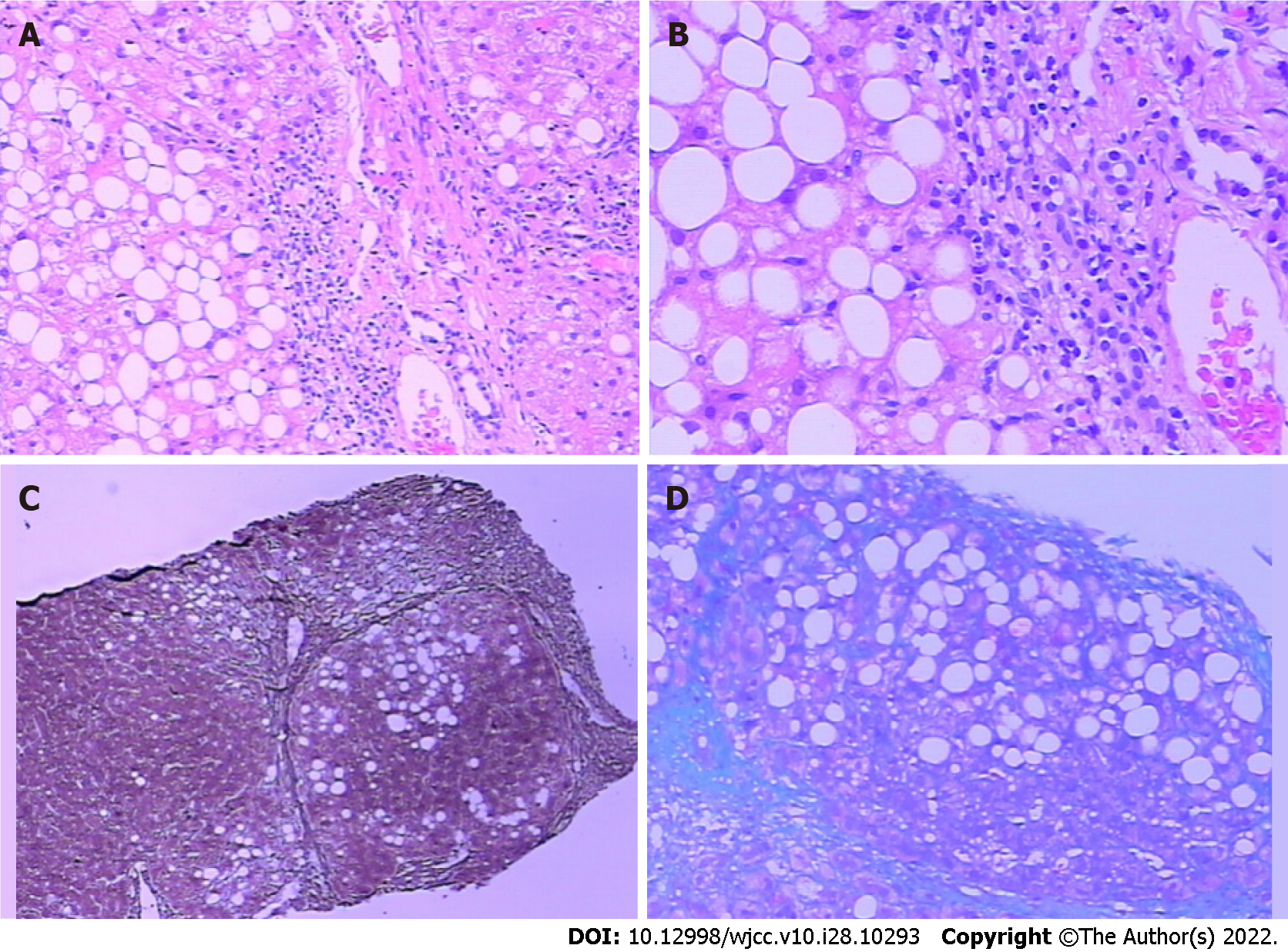
Figure 2 Liver puncture tissue and immunohistochemical staining results.
A: Periodic acid-Schiff; B: D-periodic acid-Schiff; C: Reticulation; D: Masson.
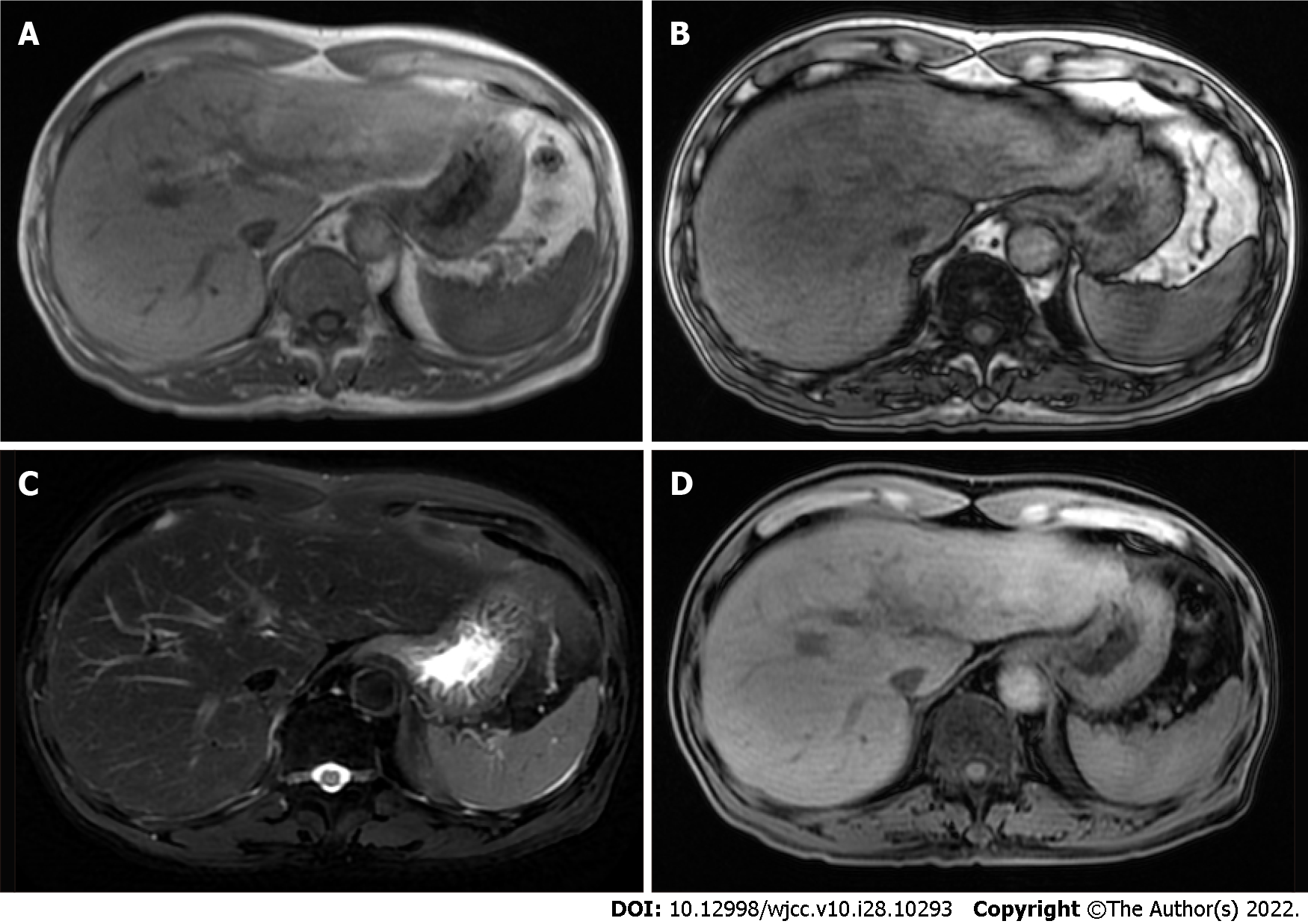
Figure 3 Plain scan and enhanced magnetic resonance imaging images of upper abdomen.
A: Positive phase; B: Reverse phase; C: Fat suppression on T2 weighted imaging; D: T1 weighted imaging.
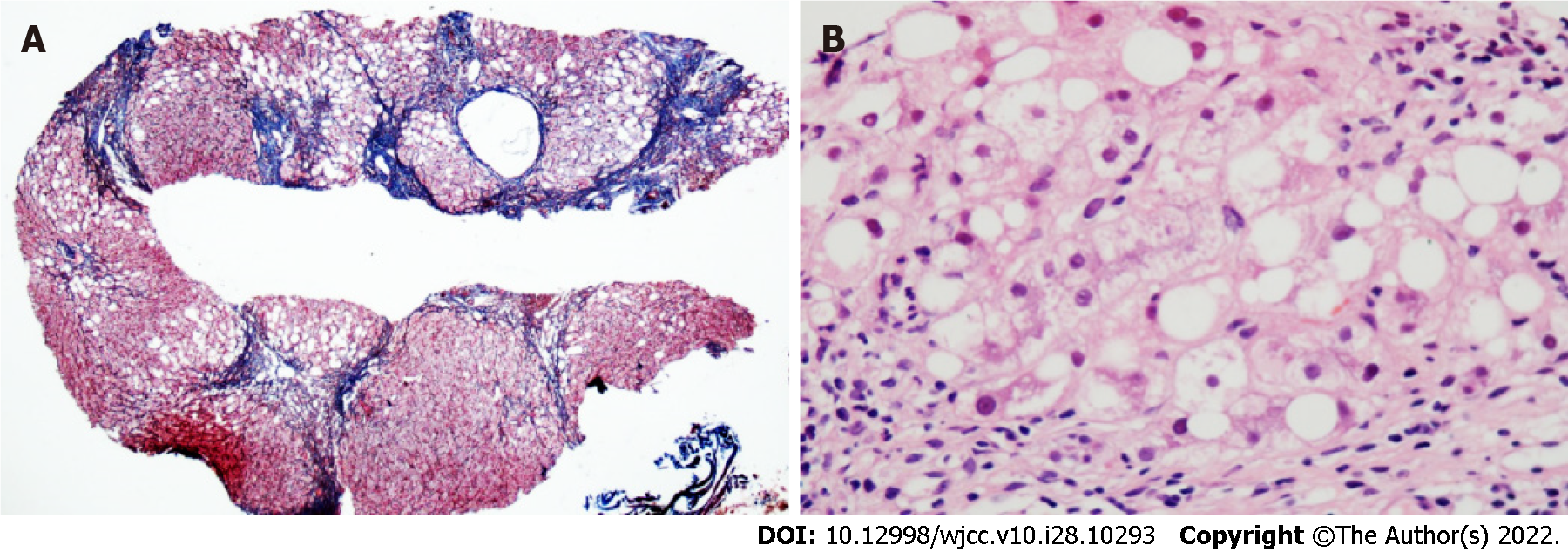
Figure 4 Liver puncture tissue and immunohistochemical staining results.
A total of 19 small and medium-sized portal areas with inflammation were observed in the tissue section. Most of the hepatocytes in the separated liver parenchyma showed vesicular steatosis, mainly distributed in about 60% of the portal area and around the necrotic zone, which were accompanied by extensive perisinusoidal fibrosis. A: Reticulation + Masson; B: Hematoxylin and eosin staining.
- Citation: Nong YB, Huang HN, Huang JJ, Du YQ, Song WX, Mao DW, Zhong YX, Zhu RH, Xiao XY, Zhong RX. Rare leptin in non-alcoholic fatty liver cirrhosis: A case report. World J Clin Cases 2022; 10(28): 10293-10300
- URL: https://www.wjgnet.com/2307-8960/full/v10/i28/10293.htm
- DOI: https://dx.doi.org/10.12998/wjcc.v10.i28.10293


