Copyright
©The Author(s) 2022.
World J Clin Cases. Oct 6, 2022; 10(28): 10286-10292
Published online Oct 6, 2022. doi: 10.12998/wjcc.v10.i28.10286
Published online Oct 6, 2022. doi: 10.12998/wjcc.v10.i28.10286
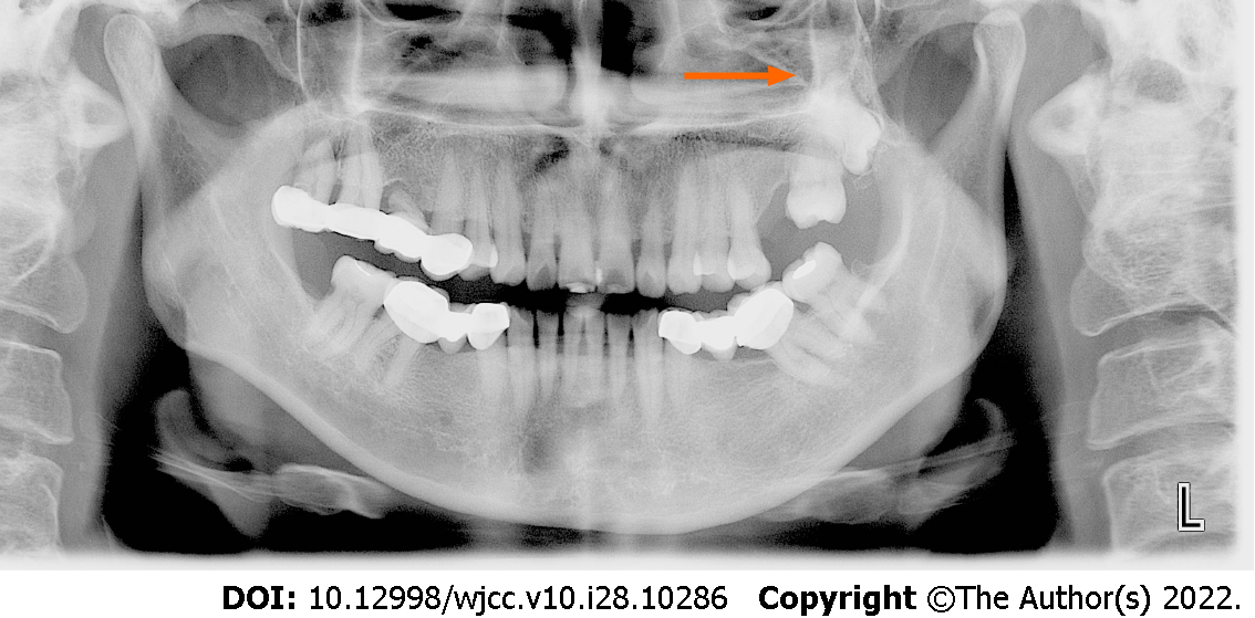
Figure 1
Panoramic film showing overlapping of the two molars without an obvious dividing line (The arrow in the figure indicates the unclear boundary between the two teeth).
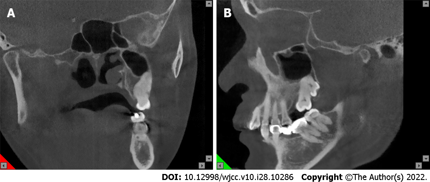
Figure 2 Coronal plane.
A: Sagittal plane; B: CBCT images. CBCT: Cone-beam computed tomography.
Figure 3 Image of extracted concrescent left maxillary second and third molars.
A-C: General views of isolated teeth (arrows in B and C indicate the junction of the two teeth).
Figure 4 Histological observation of the concrescent teeth (hematoxylin-eosin staining).
A: Scale = 1:1. B: Magnification = 4 x.
- Citation: Su J, Shao LM, Wang LC, He LJ, Pu YL, Li YB, Zhang WY. Concrescence of maxillary second molar and impacted third molar: A case report. World J Clin Cases 2022; 10(28): 10286-10292
- URL: https://www.wjgnet.com/2307-8960/full/v10/i28/10286.htm
- DOI: https://dx.doi.org/10.12998/wjcc.v10.i28.10286


