Copyright
©The Author(s) 2022.
World J Clin Cases. Oct 6, 2022; 10(28): 10279-10285
Published online Oct 6, 2022. doi: 10.12998/wjcc.v10.i28.10279
Published online Oct 6, 2022. doi: 10.12998/wjcc.v10.i28.10279
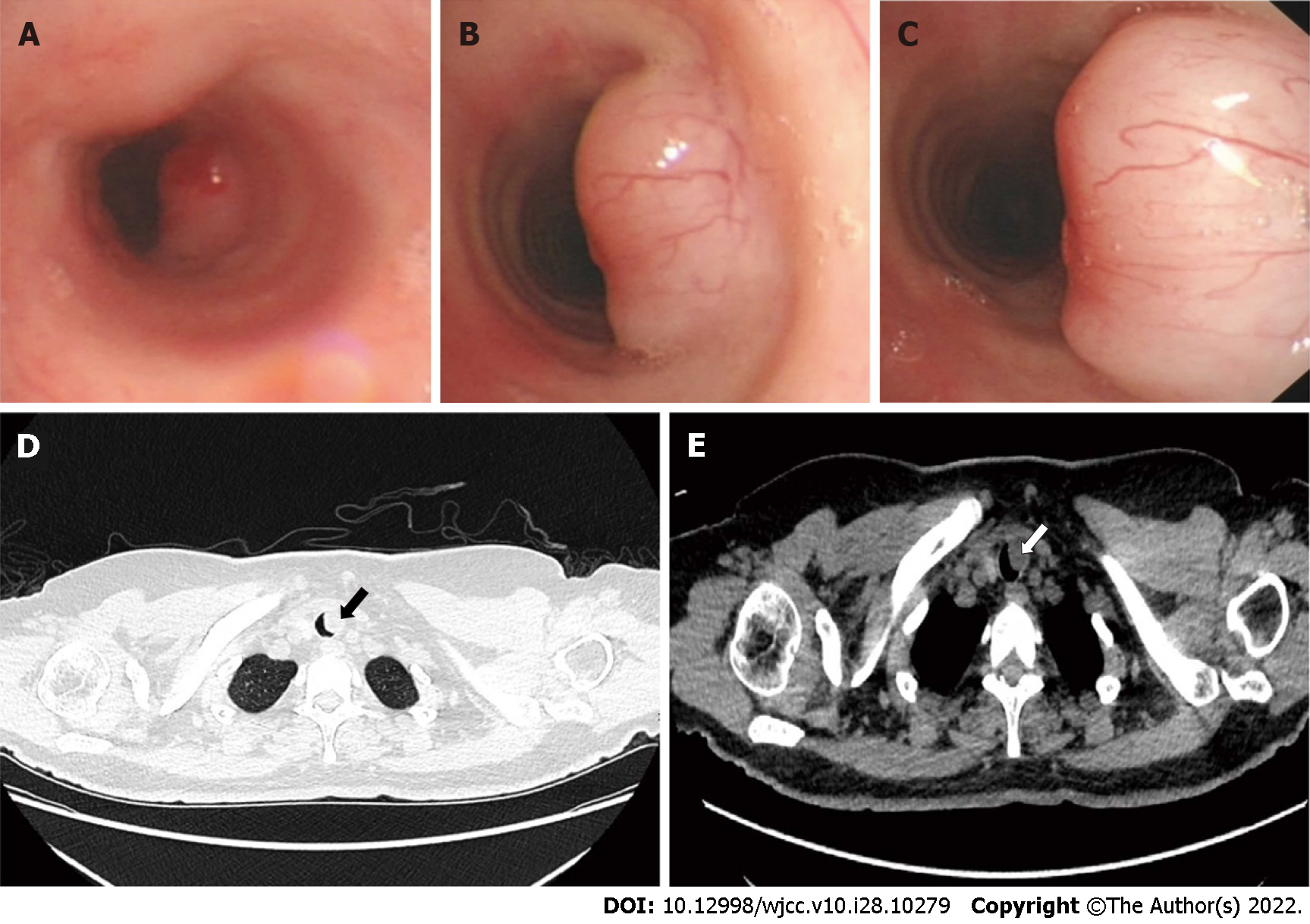
Figure 1 Bronchoscopic view and computed tomography scan of the chest showing the tracheal tumor.
A-C: Three different observation positions were observed during bronchoscopy: Distant (A), middle (B), near (C); D: Computed tomography (CT) scan, lung window; E: CT scan, mediastinal window.
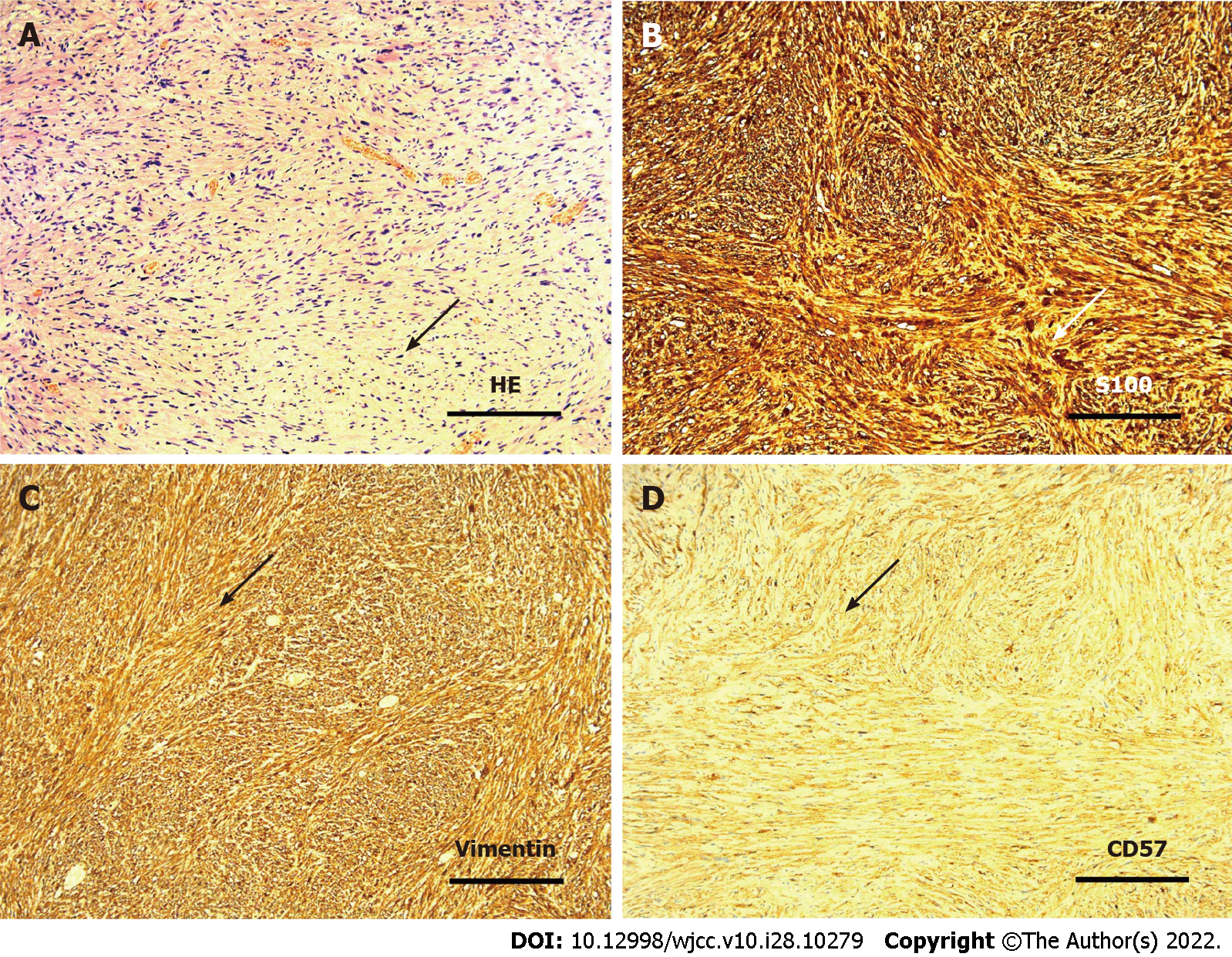
Figure 2 Results of hematoxylin and eosin staining and immunohistochemistry of tumors.
A: Photomicrograph of hematoxylin and eosin staining; B: Photomicrograph of S100 staining; C: Photomicrograph of Vimentin staining; D: Photomicrograph of CD57 staining.
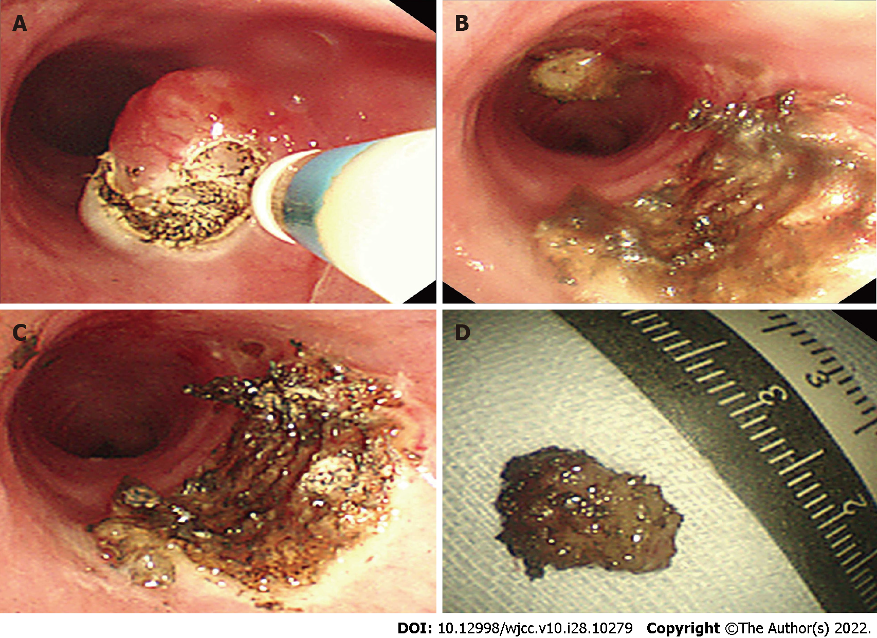
Figure 3 The process of tumor resection with a high-frequency electric knife.
A-C: Diagram of surgery; D: Resected tumor specimen.
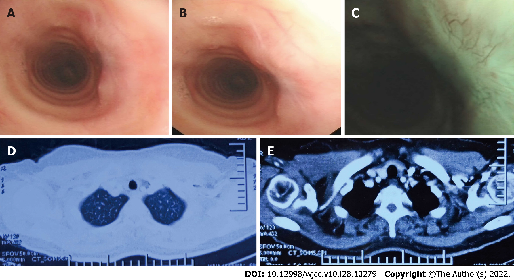
Figure 4 Postoperative re-examination.
A-C: Bronchoscopic view; D and E: Computed tomography scan of the chest.
- Citation: Shen YS, Tian XD, Pan Y, Li H. Treatment of primary tracheal schwannoma with endoscopic resection: A case report. World J Clin Cases 2022; 10(28): 10279-10285
- URL: https://www.wjgnet.com/2307-8960/full/v10/i28/10279.htm
- DOI: https://dx.doi.org/10.12998/wjcc.v10.i28.10279


