Copyright
©The Author(s) 2022.
World J Clin Cases. Oct 6, 2022; 10(28): 10244-10251
Published online Oct 6, 2022. doi: 10.12998/wjcc.v10.i28.10244
Published online Oct 6, 2022. doi: 10.12998/wjcc.v10.i28.10244
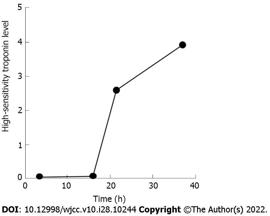
Figure 1 Troponin level.
The troponin level increased from 0.05 ng/mL to 3.92 ng/mL at admission.
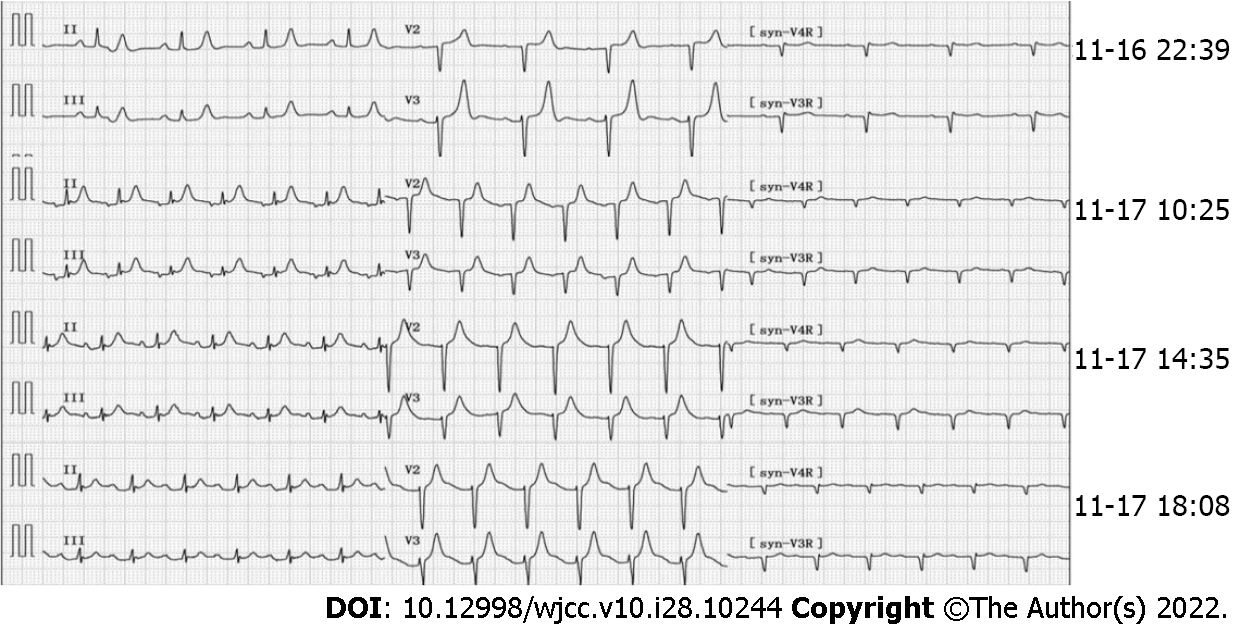
Figure 2 Electrocardiogram findings at different time points.
The initial electrocardiogram (ECG) (November 16, 2021, 22:39): Sinus rhythm, high and sharp T wave; R wave: V1-V4 progression was poor, V1 was Qr type. ECG (November 17, 2021, 10:25): Sinus tachycardia, paroxysmal atrial tachycardia, atrial fusion waves, and ST segment elevation in leads II, III, and avF was 0.10mV. ECGs (November 17, 2021, 14:35 and 18:08) reported ST-segment elevation of II, III, and avF exceeding 0.05 mV.
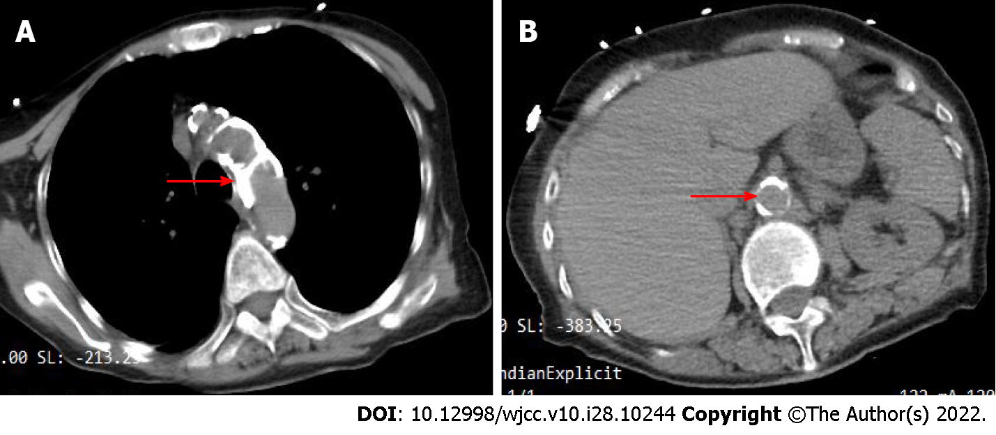
Figure 3 Abdominal computed tomography findings.
Chest and abdominal computed tomography images of the patient at admission when the patient was admitted to the hospital (November 16, 2021). A: The patient's aortic arch has markedly extensive calcification (red arrow); B: The patient's abdominal aorta has obvious calcification (red arrow indicates the opening of the superior mesenteric artery).
Figure 4 Percutaneous coronary intervention findings.
No significant abnormalities were found in the left and right coronary arteries (November 17, 2021). A: Left anterior descending coronary artery (red arrow); B: Left circumflex coronary artery (red arrow); C: Right coronary artery (red arrow).
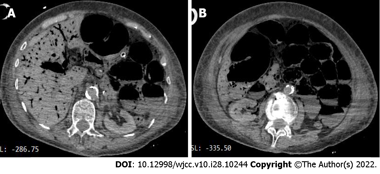
Figure 5 Abdominal computed tomography findings.
Extensive gas and fluid in the small intestine, multiple liquid and gas plane shadows could be seen, and the gas was accumulated in the intestinal wall and the corresponding mesangial blood vessels of the hepatic portal system, suggesting the possibility of extensive necrosis of the intestinal wall. A: A lot of gas in the hepatic portal system; B: Extensive gas and fluid in the small intestine.
- Citation: Ding P, Zhou Y, Long KL, Zhang S, Gao PY. Acute mesenteric ischemia due to percutaneous coronary intervention: A case report. World J Clin Cases 2022; 10(28): 10244-10251
- URL: https://www.wjgnet.com/2307-8960/full/v10/i28/10244.htm
- DOI: https://dx.doi.org/10.12998/wjcc.v10.i28.10244


