Copyright
©The Author(s) 2022.
World J Clin Cases. Oct 6, 2022; 10(28): 10227-10235
Published online Oct 6, 2022. doi: 10.12998/wjcc.v10.i28.10227
Published online Oct 6, 2022. doi: 10.12998/wjcc.v10.i28.10227
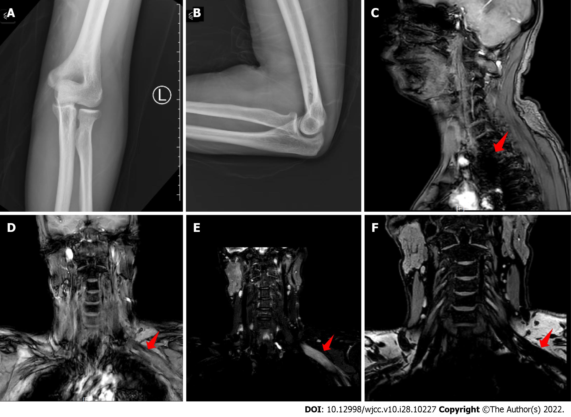
Figure 1 X-ray and magnetic resonance images.
A and B: No significant abnormality on antero-posterior (A) and lateral (B) X-rays of the left elbow joint; C and D: Sagittal (C) and coronal (D) magnetic resonance images (MRI) showing significant abnormal signal in the left brachial plexus; E and F: Coronal MRI showing significant abnormal signal in the left brachial plexus. The red arrow points to the lesion.
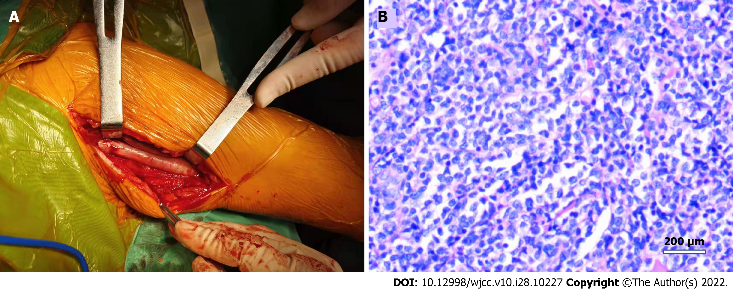
Figure 2 Intraoperative and histological images.
A: Intraoperative thickening and degeneration of the left ulnar nerve; B: Hematoxylin and eosin-stained section showing neuroepithelial and lymph node pathology.
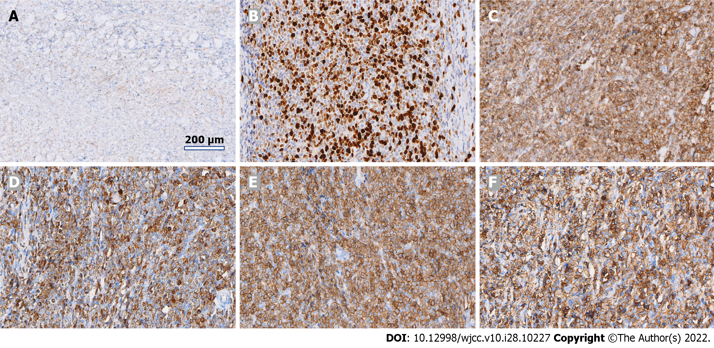
Figure 3 Histopathology of myeloid sarcoma.
Immunopathological examination shows tissue of lymphohaematopoietic lineage. A: CD21; B: Ki-67; C: LCA; D: MPO; E: CD117; F: CD34.
- Citation: Li DP, Liu CZ, Jeremy M, Li X, Wang JC, Nath Varma S, Gai TT, Tian WQ, Zou Q, Wei YM, Wang HY, Long CJ, Zhou Y. Myeloid sarcoma with ulnar nerve entrapment: A case report. World J Clin Cases 2022; 10(28): 10227-10235
- URL: https://www.wjgnet.com/2307-8960/full/v10/i28/10227.htm
- DOI: https://dx.doi.org/10.12998/wjcc.v10.i28.10227


