Copyright
©The Author(s) 2022.
World J Clin Cases. Oct 6, 2022; 10(28): 10201-10207
Published online Oct 6, 2022. doi: 10.12998/wjcc.v10.i28.10201
Published online Oct 6, 2022. doi: 10.12998/wjcc.v10.i28.10201
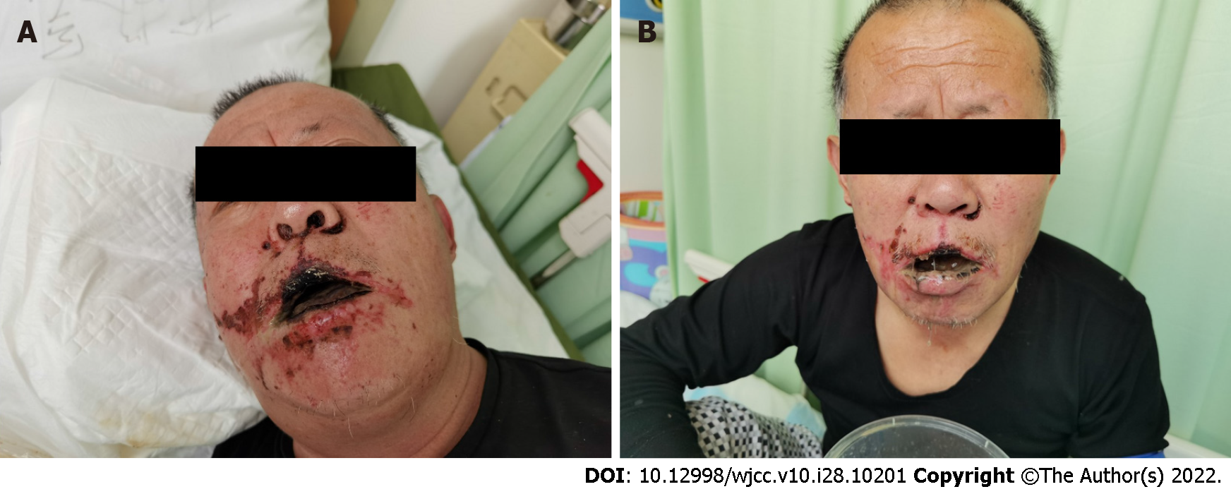
Figure 1 Comparison of facial manifestations before and after therapy.
A: Upon admission, there are scattered black burn marks on the face, especially in the nasolabial sulcus on both sides. Slightly limited mouth opening (approximately 2.5 cm). Mucous membranes, such as the lips and tongue, were black with obvious ulceration and bleeding; B: 7 d after therapy, the black burn marks on the face were less than before, and the bleeding of lip and tongue ulcer had reduced slightly. However, the mouth opening remained limited.
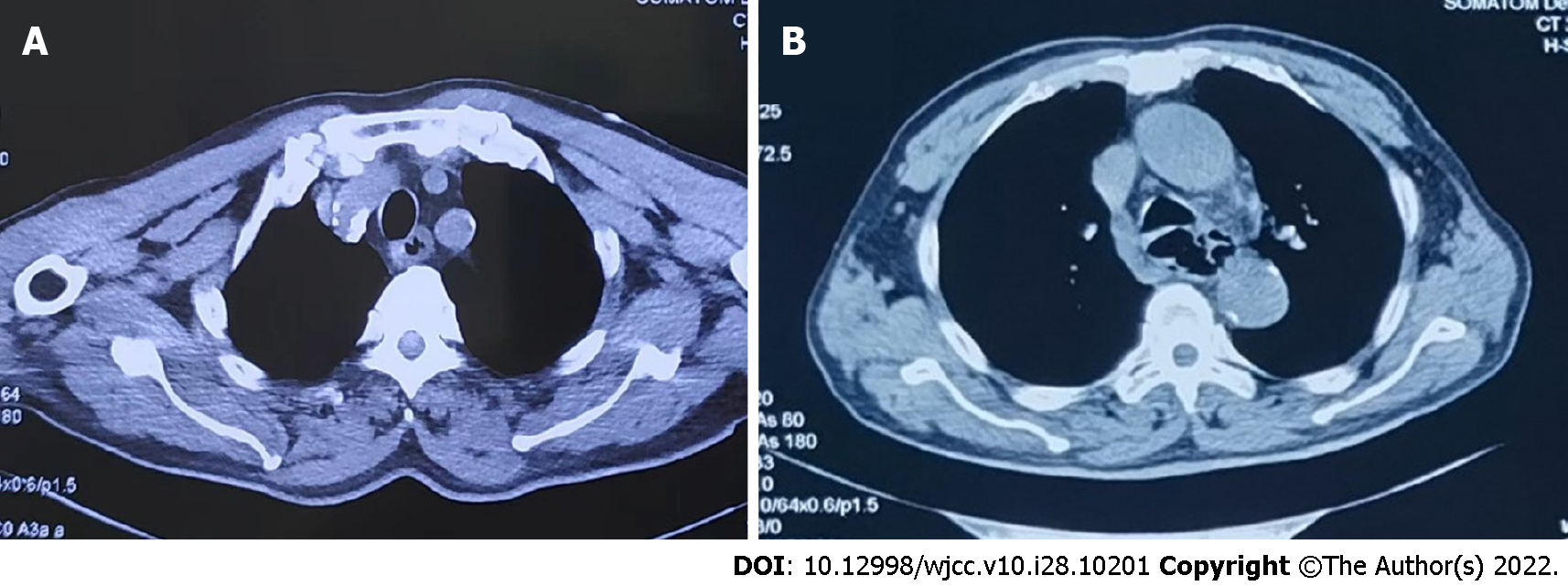
Figure 2 Computed tomography images of the lesion.
A: Upon admission showing artery calcification, thickened oesophageal wall, and a dilated lumen; B: On day 7 showing thickened, dilated, and irregularly shaped oesophageal wall.
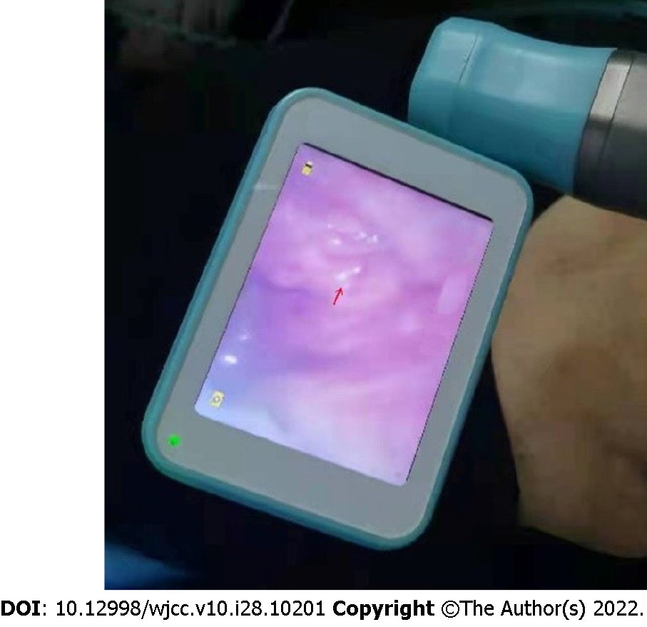
Figure 3 Laryngoscopy.
Day 12 laryngoscopy showing red granulation tissue after necrotic tissue abscission.
- Citation: Li YQ, Yu GC, Shi LK, Zhao LW, Wen ZX, Kan BT, Jian XD. Clinical analysis of pipeline dredging agent poisoning: A case report. World J Clin Cases 2022; 10(28): 10201-10207
- URL: https://www.wjgnet.com/2307-8960/full/v10/i28/10201.htm
- DOI: https://dx.doi.org/10.12998/wjcc.v10.i28.10201


