Copyright
©The Author(s) 2022.
World J Clin Cases. Oct 6, 2022; 10(28): 10193-10200
Published online Oct 6, 2022. doi: 10.12998/wjcc.v10.i28.10193
Published online Oct 6, 2022. doi: 10.12998/wjcc.v10.i28.10193
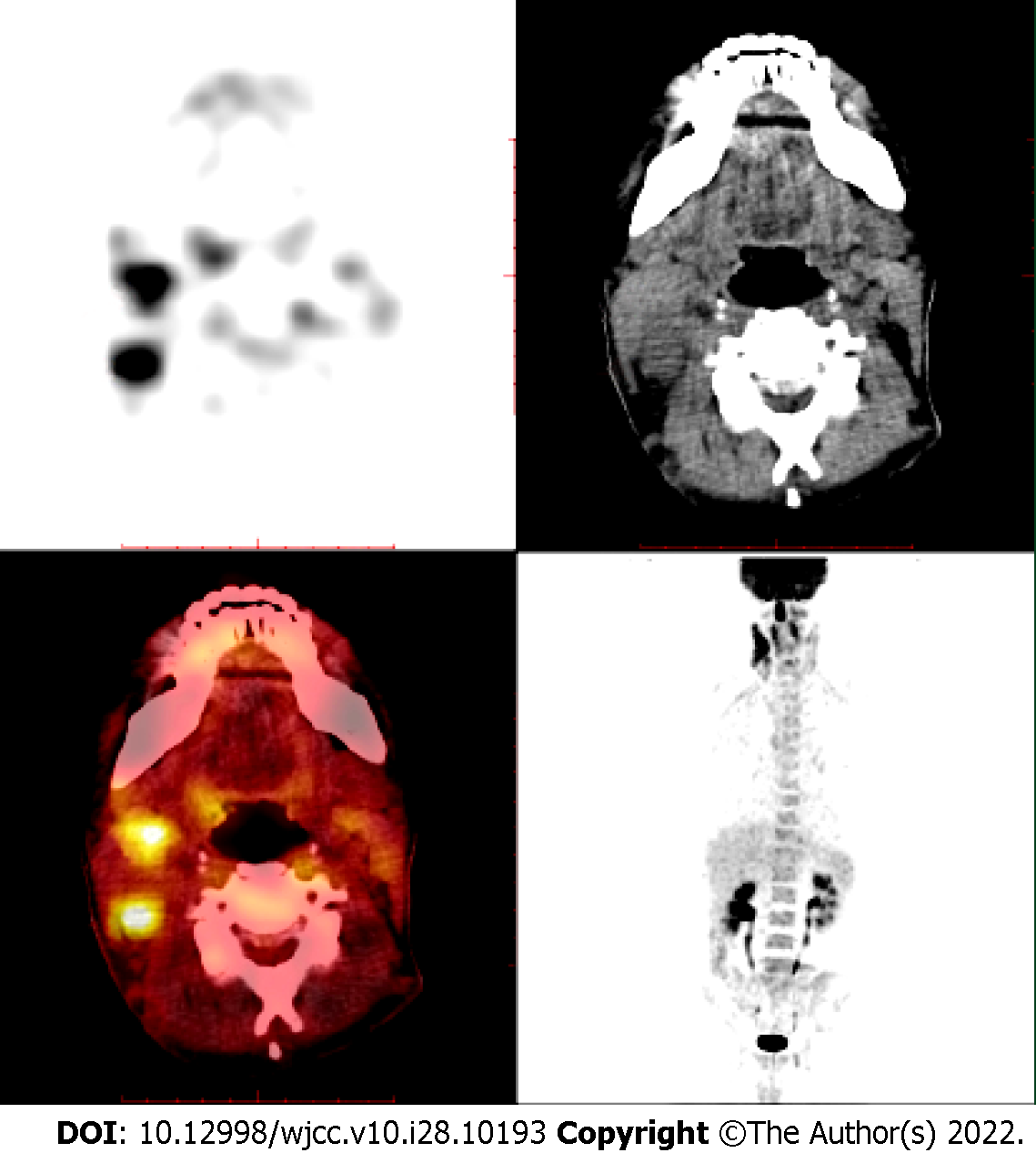
Figure 1 Multiple lymphadenopathies in the bilateral neck and supraclavicular area (especially in the right neck), strip-shaped soft tissue density shadow in the right nasal cavity, and significantly increased fluorodeoxyglucose metabolism as revealed by standard uptake value (SUV) with a Deauville score of 7.
6.
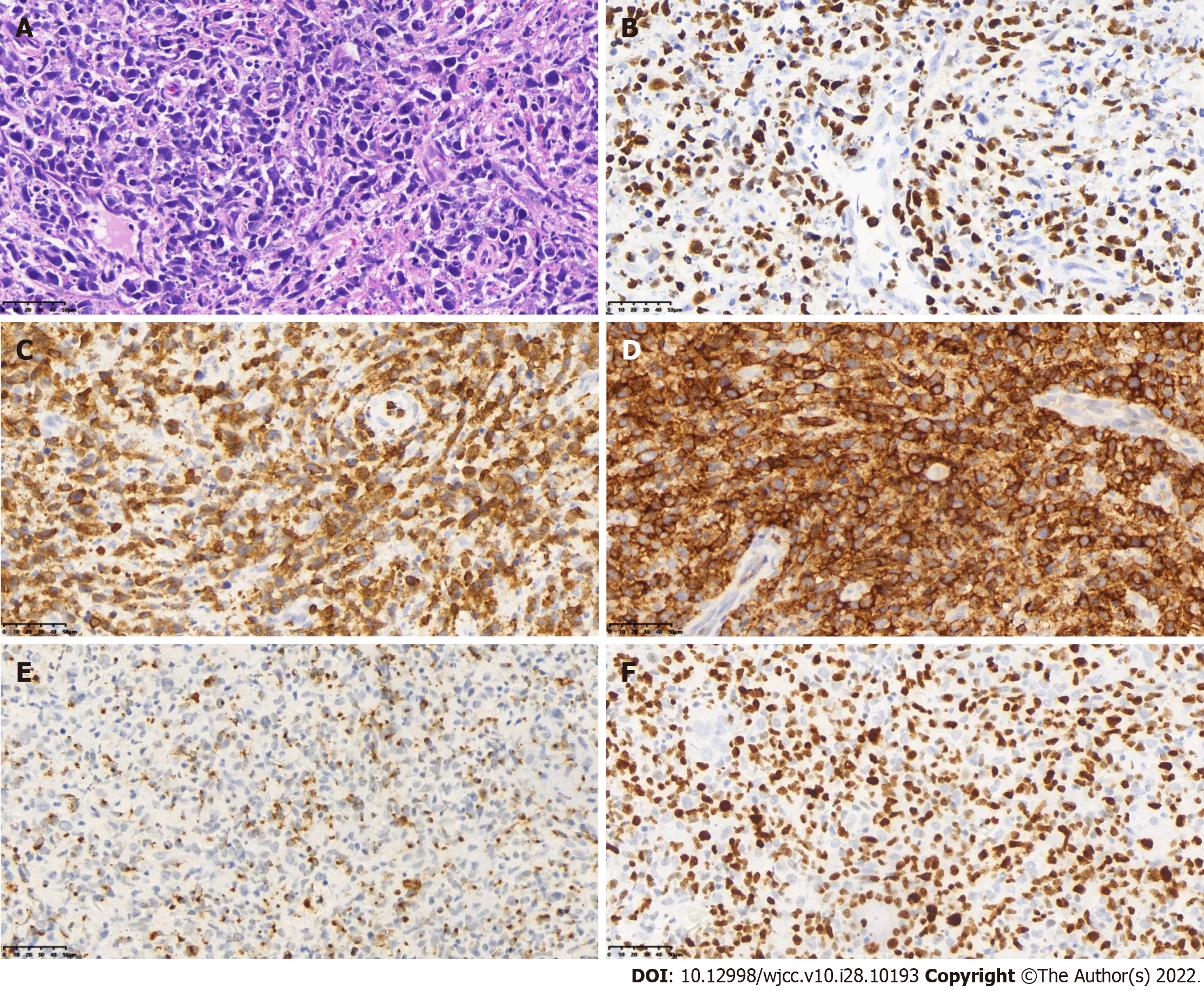
Figure 2 Molecular in situ hybridization demonstrated positivity for EBER.
A: Tumor cells are large, nucleus pleomorphic or elongated, deeply stained, diffuse, and patchy (hematoxylin-eosin staining, × 400); B: EBER positivity (in situ hybridization, × 400); C: CD3 positivity (Envision two-step immunohistochemical staining, × 400); D: CD56-positivity (Envision two-step immunohistochemical staining, × 400); E: Granzyme B positivity (Envision two-step immunohistochemical staining, × 400); F: Ki-67 positive expression rate of 80%.
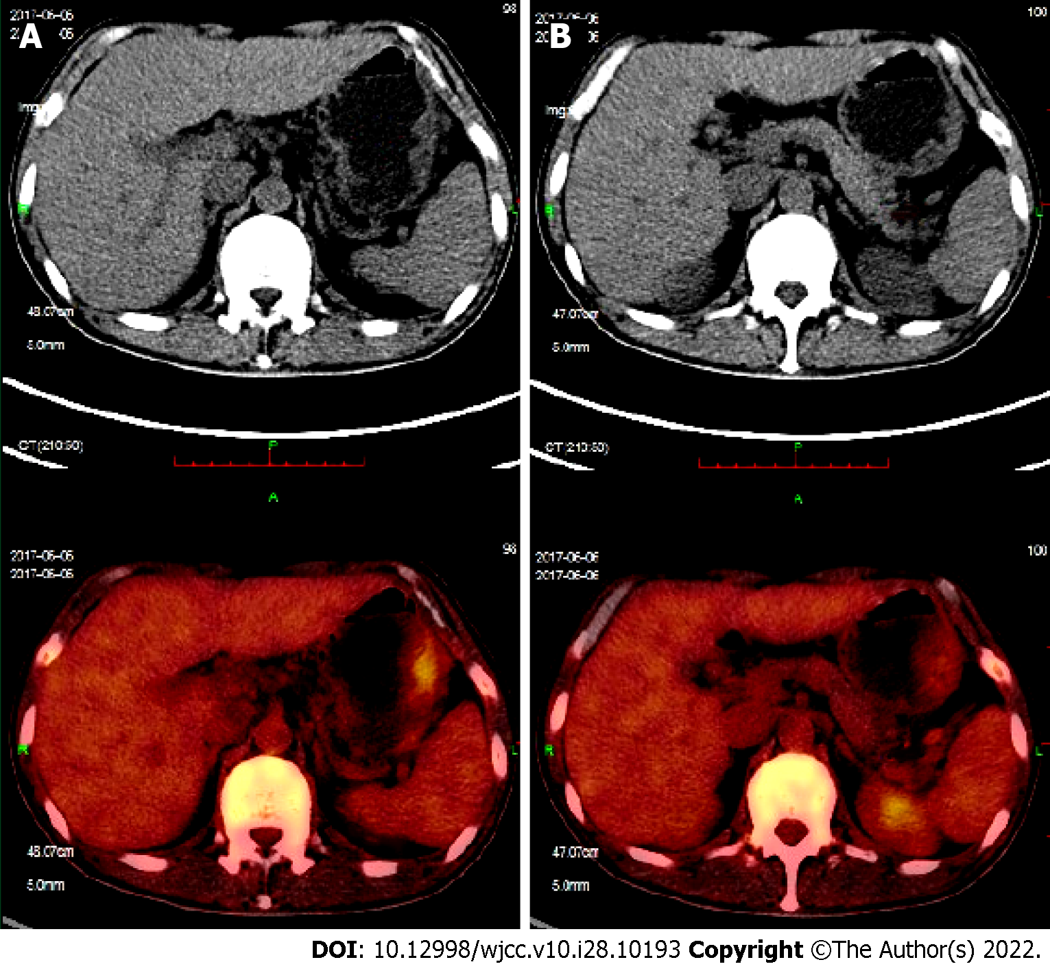
Figure 3 Positron emission tomography/computed tomography images.
A and B: New large-curved wall thickening of the gastric body, multiple nodules of the spleen, and multiple lymphadenopathies around the stomach.
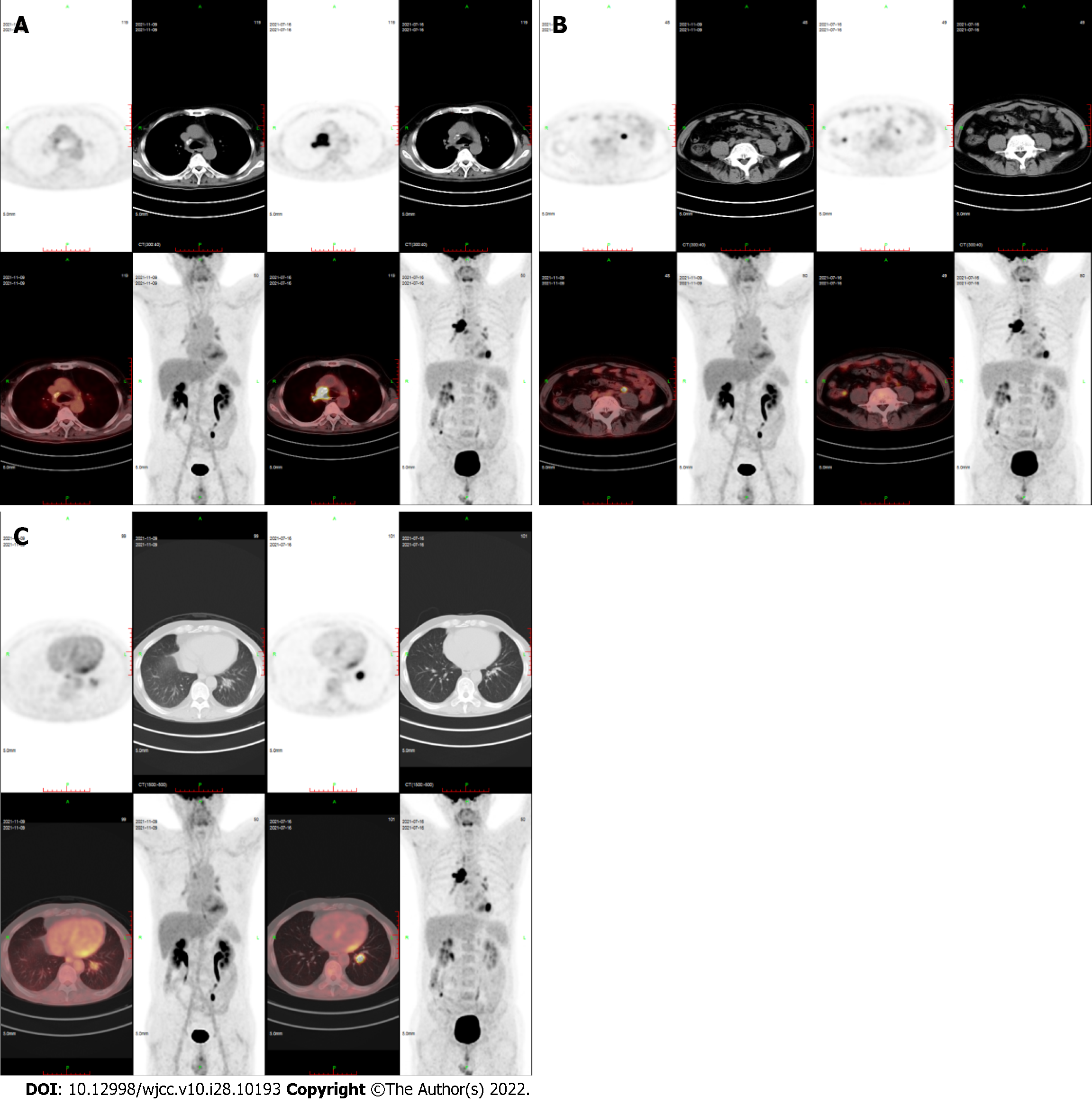
Figure 4 Positron emission tomography/computed tomography images.
A-C: Compared to the film on July 16, 2021, that taken on November 9, 2021 showed that right mediastinal paratracheal, right main bronchial, subcarinal lymph nodes and ileocecal small nodules had subsided and become inactive, and the metabolism of left lower lobe nodules was decreased significantly (SUVmax decreased from 10.2 to 3.8).
- Citation: Li LJ, Zhang JY. Treatment of refractory/relapsed extranodal NK/T cell lymphoma with decitabine plus anti-PD-1: A case report. World J Clin Cases 2022; 10(28): 10193-10200
- URL: https://www.wjgnet.com/2307-8960/full/v10/i28/10193.htm
- DOI: https://dx.doi.org/10.12998/wjcc.v10.i28.10193


