Copyright
©The Author(s) 2022.
World J Clin Cases. Oct 6, 2022; 10(28): 10180-10185
Published online Oct 6, 2022. doi: 10.12998/wjcc.v10.i28.10180
Published online Oct 6, 2022. doi: 10.12998/wjcc.v10.i28.10180
Figure 1 Tumor enhancement and metabolic imaging.
A-C: Multiphase MR enhanced images; D-E: Positron emission tomography/computed tomography images, and F: Pathological sections of tumor (HE × 300).
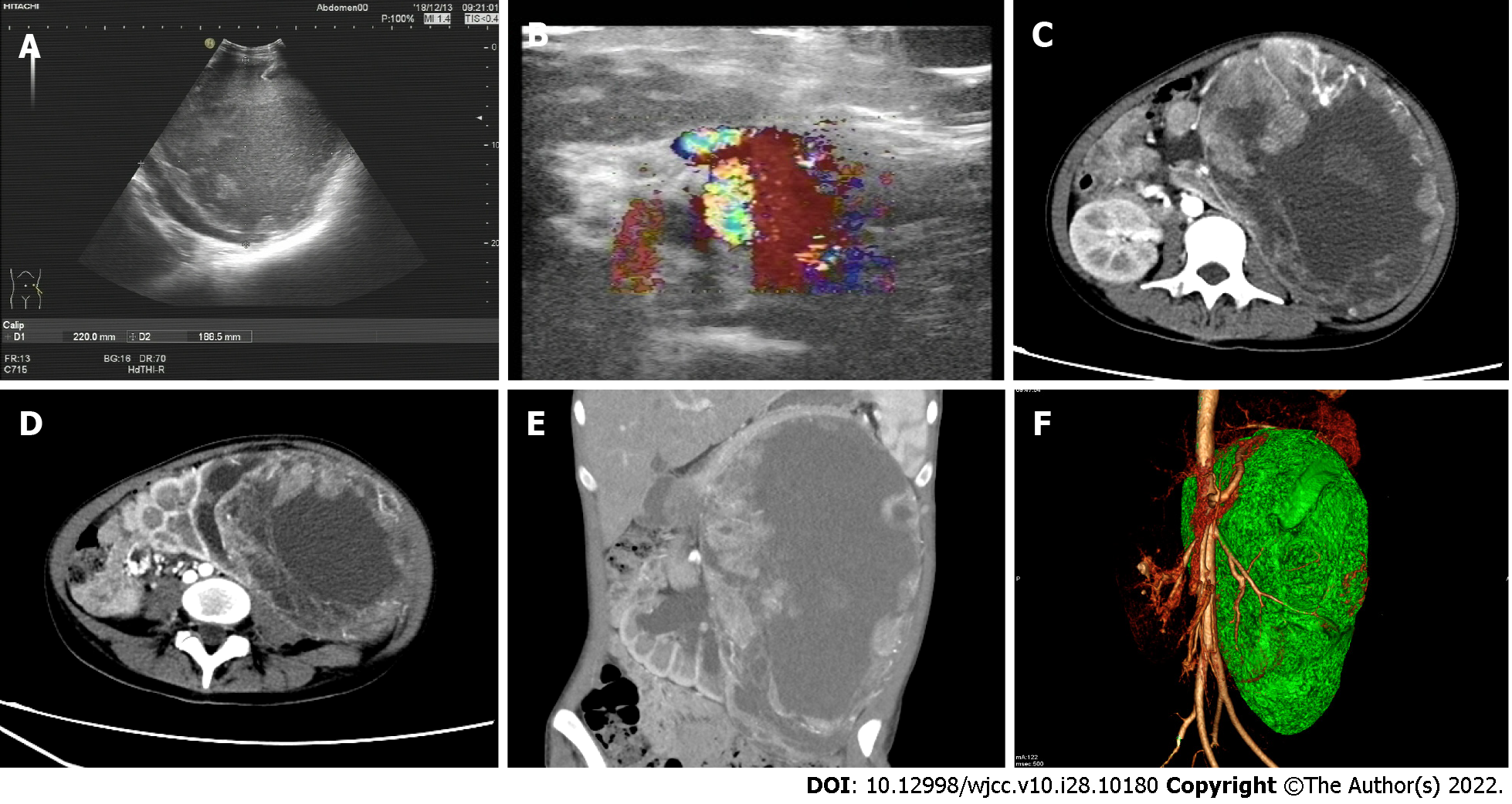
Figure 2 Tumor blood supply and vascular imaging.
A and B: Ultrasonography and color Doppler flow imaging; C-E: Multiphase computed tomography enhanced images; F: Vascular and tumor volume reconstruction.
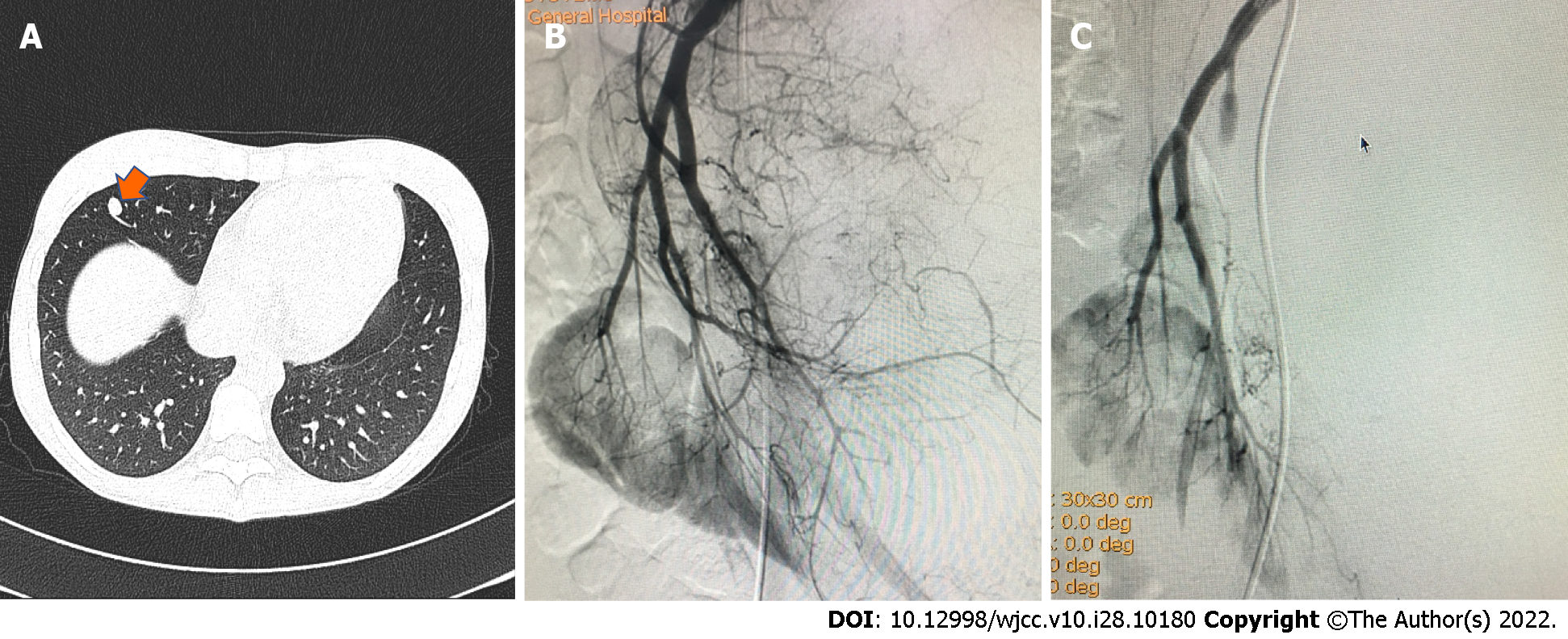
Figure 3 Tumor metastasis and transarterial chemoembolization images.
A: Metastases in the middle lobe of the right lung (orange arrow); B: DSA showed tumor vascular mass staining; C: Digital subtraction angiography showed disappearance of tumor vascular mass staining after transarterial chemoembolization (black arrow).
- Citation: Wang P, Zhang X, Shao SH, Wu F, Du FZ, Zhang JF, Zuo ZW, Jiang R. Chemotherapy, transarterial chemoembolization, and nephrectomy combined treated one giant renal cell carcinoma (T3aN1M1) associated with Xp11.2/TFE3: A case report. World J Clin Cases 2022; 10(28): 10180-10185
- URL: https://www.wjgnet.com/2307-8960/full/v10/i28/10180.htm
- DOI: https://dx.doi.org/10.12998/wjcc.v10.i28.10180


