Copyright
©The Author(s) 2022.
World J Clin Cases. Oct 6, 2022; 10(28): 10146-10154
Published online Oct 6, 2022. doi: 10.12998/wjcc.v10.i28.10146
Published online Oct 6, 2022. doi: 10.12998/wjcc.v10.i28.10146
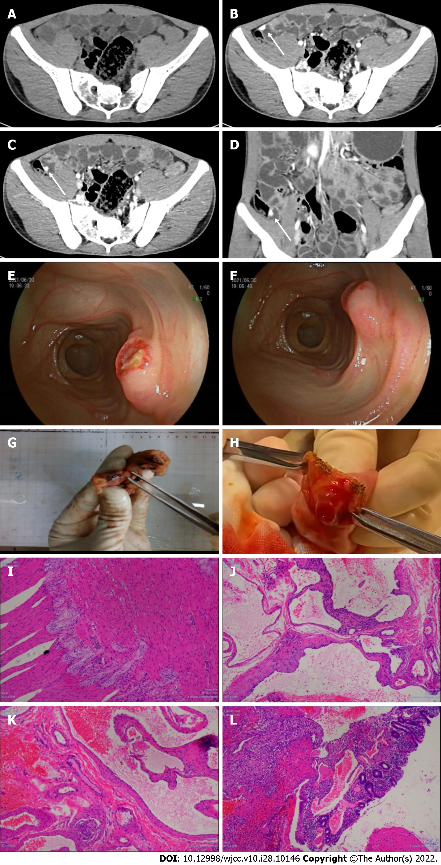
Figure 1 Imaging, enteroscopy, surgery and pathology of the ileal cavernous hemangioma.
A: No obvious lesion was found in the unenhanced phase; B: A high-density shadow was found in the arterial phase; C and D: A high-density shadow was found in the venous phase; E and F: The transanal double-balloon enteroscopy reached the upper segment of the ileum. In the middle and lower segments of the ileum, the mucosa was rough and eroded, and the villi were absent. A submucosal bulge of about 1.2 cm × 1.0 cm was observed approximately 340 cm proximal to the ileocecal valve, where the ulcers developed. The surrounding mucosa was flushed, presented edema, hard in texture, and easily bleeding, with many thickened blood vessels supplying the lesion; G and H: Gross specimen of the ileal cavernous hemangioma; I-L: Hematoxylin–eosin staining showing a blood-filled sinus-like space in the ileum submucosa; I: Original magnification 20 ×; J-L: Original magnification 10 ×.
- Citation: Yao L, Li LW, Yu B, Meng XD, Liu SQ, Xie LH, Wei RF, Liang J, Ruan HQ, Zou J, Huang JA. Cavernous hemangioma of the ileum in a young man: A case report and review of literature. World J Clin Cases 2022; 10(28): 10146-10154
- URL: https://www.wjgnet.com/2307-8960/full/v10/i28/10146.htm
- DOI: https://dx.doi.org/10.12998/wjcc.v10.i28.10146


