Copyright
©The Author(s) 2022.
World J Clin Cases. Oct 6, 2022; 10(28): 10120-10129
Published online Oct 6, 2022. doi: 10.12998/wjcc.v10.i28.10120
Published online Oct 6, 2022. doi: 10.12998/wjcc.v10.i28.10120
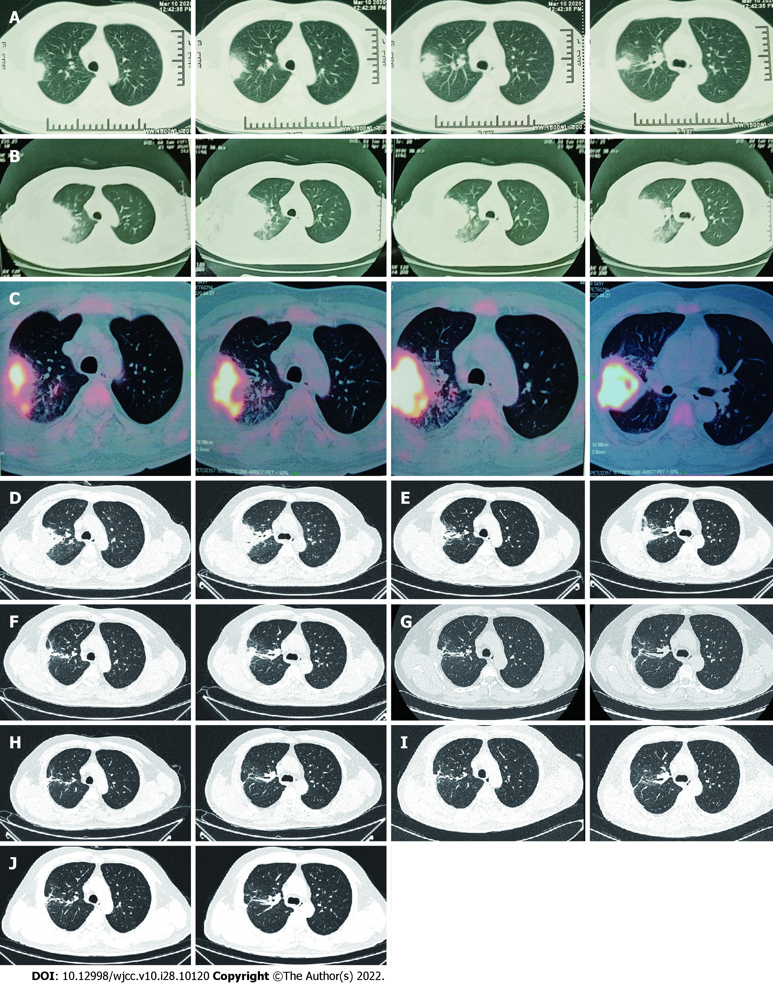
Figure 1 Computed tomography of right lung.
A: The first scan demonstrated a mass and atelectasis in the right upper lobe on March 20, 2020; B: Chest Computed Tomography (CT) showed aggravation of the original lesion on April 23, 2020; C: Positron emission tomography-CT showed that the mass lesions in the posterior segment of the right upper lobe had significantly increased glucose metabolism inhomogeneously on April 27, 2020; D: Primary lesion of the right upper lobe reduced on May 23, 2020; E-J: Repeated pulmonary CT showed primary lesion size of the right upper lobe continued to decrease on different times, involving June 22, 2020 (E), July 29, 2020 (F), September 8, 2020 (G), November 20, 2020 (H), January 7, 2021 (I) and March 25, 2021 (J). CT: Computed tomography.
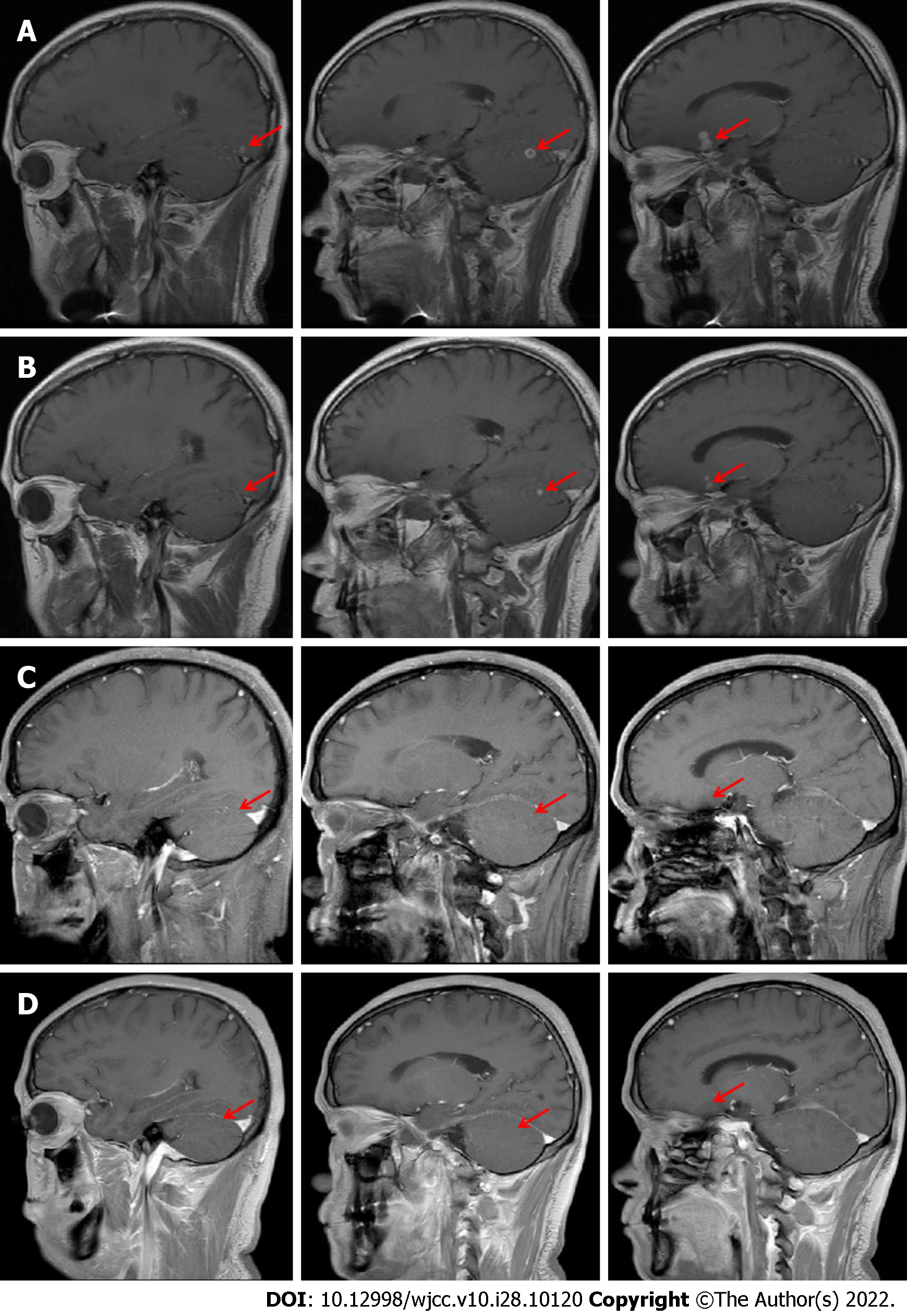
Figure 2 Brain magnetic resonance imaging findings.
A: Cranial magnetic resonance imaging (MRI) showed multiple lesions in the left occipital lobe, right cerebellar hemisphere and left frontal lobe on May 13, 2020; B: Cranial MRI showed a decrease of the original lesion on June 15, 2020; C: Primary lesions of the brain maintained shrinkage on July 31, 2020; D: Primary lesions of the brain almost completely disappeared on September 10, 2020. MRI: Magnetic resonance imaging.
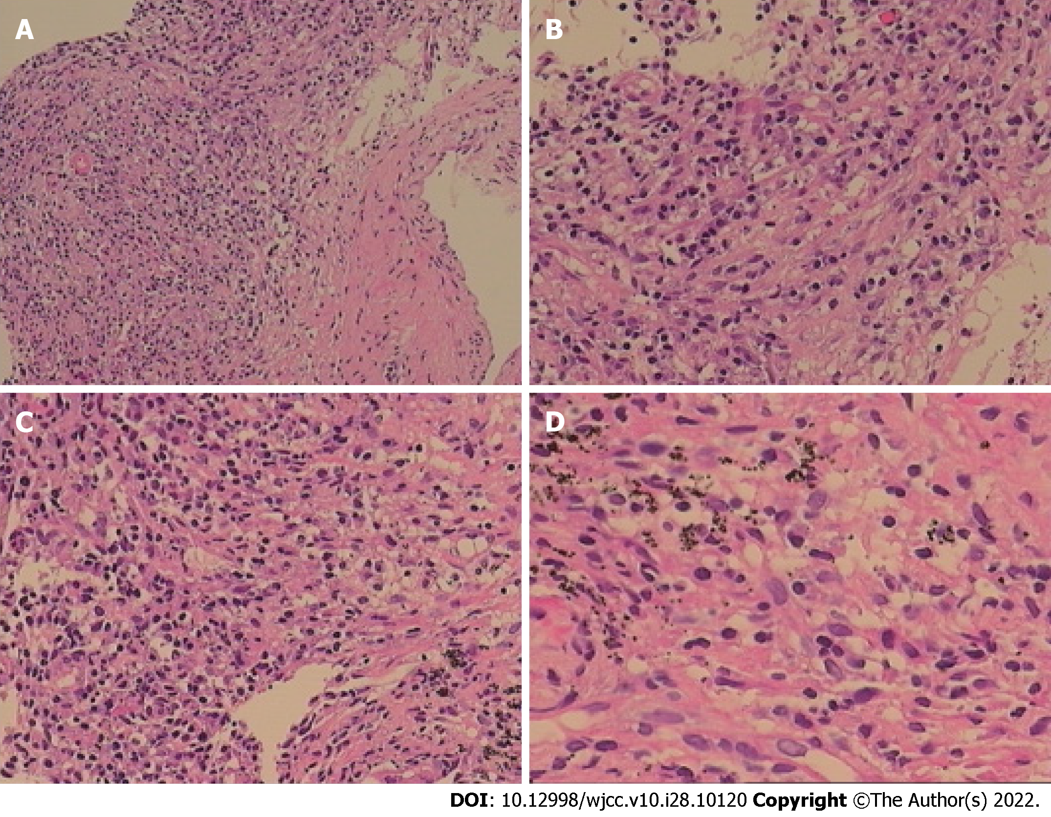
Figure 3 Chronic inflammation with acute activity and fibrous hyperplasia, with focal necrosis in the right upper lobe.
A: A large number of chronic inflammatory cells with a small amount of neutrophil infiltration in hyperplastic fibrous connective tissue (Original magnification: 100 ×; scale bar: 100 μm); B: There are collagen hyperplasia and no obvious alveolar epithelial cells in some areas (Original magnification: 200 ×; scale bar: 100 μm); C and D: In addition, there are phagocytic cells that engulf carbonic acid in some areas, inflammatory cell infiltration and capillary endothelial cells in the interstitium (C: 200 ×; scale bar: 100 μm; D: 400 ×; scale bar: 100 μm).
- Citation: Li T, Chen YX, Lin JJ, Lin WX, Zhang WZ, Dong HM, Cai SX, Meng Y. Successful treatment of disseminated nocardiosis diagnosed by metagenomic next-generation sequencing: A case report and review of literature. World J Clin Cases 2022; 10(28): 10120-10129
- URL: https://www.wjgnet.com/2307-8960/full/v10/i28/10120.htm
- DOI: https://dx.doi.org/10.12998/wjcc.v10.i28.10120


