Copyright
©The Author(s) 2022.
World J Clin Cases. Oct 6, 2022; 10(28): 10031-10041
Published online Oct 6, 2022. doi: 10.12998/wjcc.v10.i28.10031
Published online Oct 6, 2022. doi: 10.12998/wjcc.v10.i28.10031
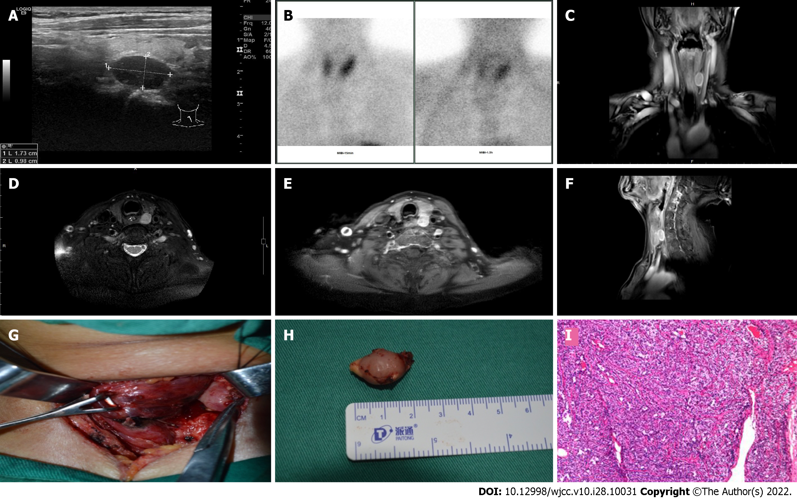
Figure 1 Parathyroid adenoma on the dorsal of the superior pole of the left lobe of the thyroid gland.
A: Ultrasonography shows a hypoechoic nodule on the back of the left lobe of the thyroid gland; B: The nodule was positive on technetium-99 m sestamibi single-photon emission computed tomography/computed tomography; C-F: Magnetic resonance imaging with contrast shows the nodule and is distinct in the coronal (C), axial (D and E), and sagittal positions (F), respectively; G: The correlation between parathyroid adenoma (PA) and the left thyroid lobe is shown, and the PA capsule should be carefully protected during surgery; H: The size of the PA was measured; I: The pathological result confirmed that the nodule was a PA.
- Citation: Peng ZX, Qin Y, Bai J, Yin JS, Wei BJ. Analysis of the successful clinical treatment of 140 patients with parathyroid adenoma: A retrospective study. World J Clin Cases 2022; 10(28): 10031-10041
- URL: https://www.wjgnet.com/2307-8960/full/v10/i28/10031.htm
- DOI: https://dx.doi.org/10.12998/wjcc.v10.i28.10031


