Copyright
©The Author(s) 2022.
World J Clin Cases. Sep 16, 2022; 10(26): 9518-9523
Published online Sep 16, 2022. doi: 10.12998/wjcc.v10.i26.9518
Published online Sep 16, 2022. doi: 10.12998/wjcc.v10.i26.9518
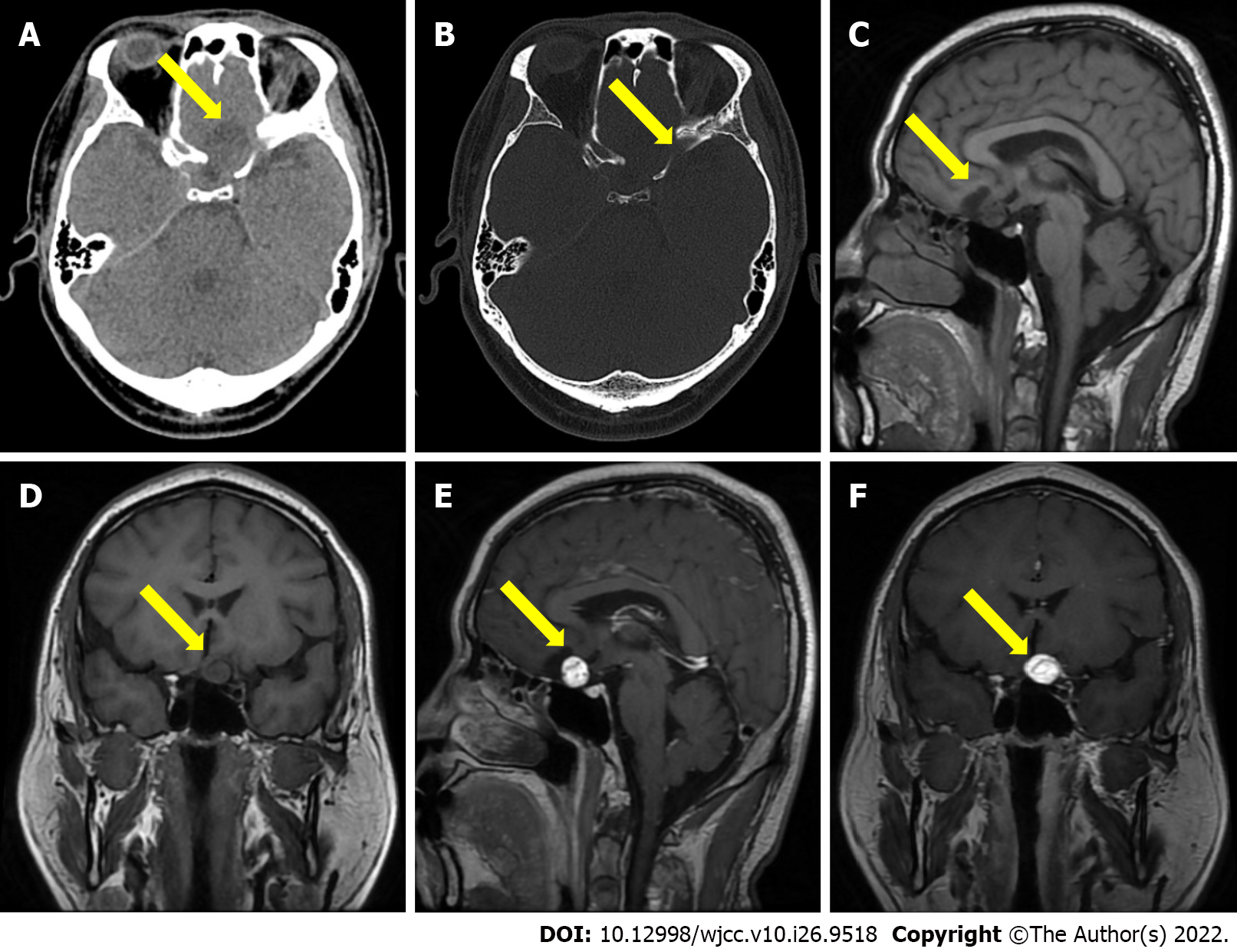
Figure 1 Brain computed tomography and magnetic resonance imaging scan findings.
A and B: The computed tomography imaging showing a quasi circle-shaped and well-defined 24.0 mm diameter lesion with hybrid density in the anterior skull base; C and D: Sagittal and coronal T1-weighted image showing the lesion has both slightly low and low signal; E and F: Sagittal and coronal gadolinium-enhanced T1-weighted image showing the obviously enhancement of the mural nodule and flow void in the mass lesion.
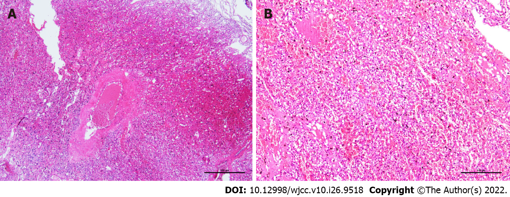
Figure 2 Postoperative pathological findings.
A: Histopathological examination showing an abundant capillary network and foamy stromal cells, and the nucleus of these cells exhibited slight atypia, hematoxylin and eosin staining (HE), 4 ×; B: HE, 10 ×.
- Citation: Xu ST, Cao X, Yin XY, Zhang JY, Nan J, Zhang J. Supratentorial hemangioblastoma at the anterior skull base: A case report. World J Clin Cases 2022; 10(26): 9518-9523
- URL: https://www.wjgnet.com/2307-8960/full/v10/i26/9518.htm
- DOI: https://dx.doi.org/10.12998/wjcc.v10.i26.9518


