Copyright
©The Author(s) 2022.
World J Clin Cases. Sep 16, 2022; 10(26): 9462-9469
Published online Sep 16, 2022. doi: 10.12998/wjcc.v10.i26.9462
Published online Sep 16, 2022. doi: 10.12998/wjcc.v10.i26.9462
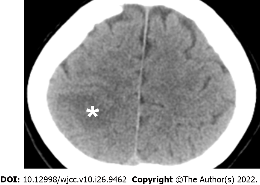
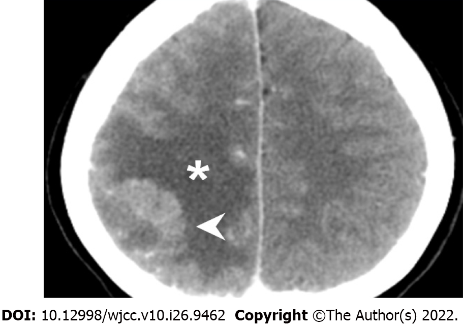
Figure 1 The initial brain computed tomography without contrast.
Asterisk: A low-density area at the right parietal lobe indicating acute ischemic stroke.
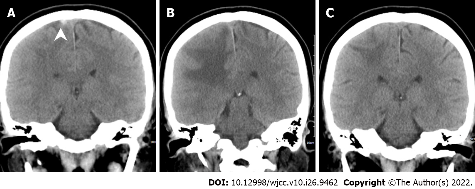
Figure 2 The serial brain computed tomography scans.
A: Day 17; B: Day 33; C: Day 61. Arrowhead: A hyperacute thrombus is found in the right cortical vein on day 17 after the vaccination. The cortical vein thrombus resolved on day 33.
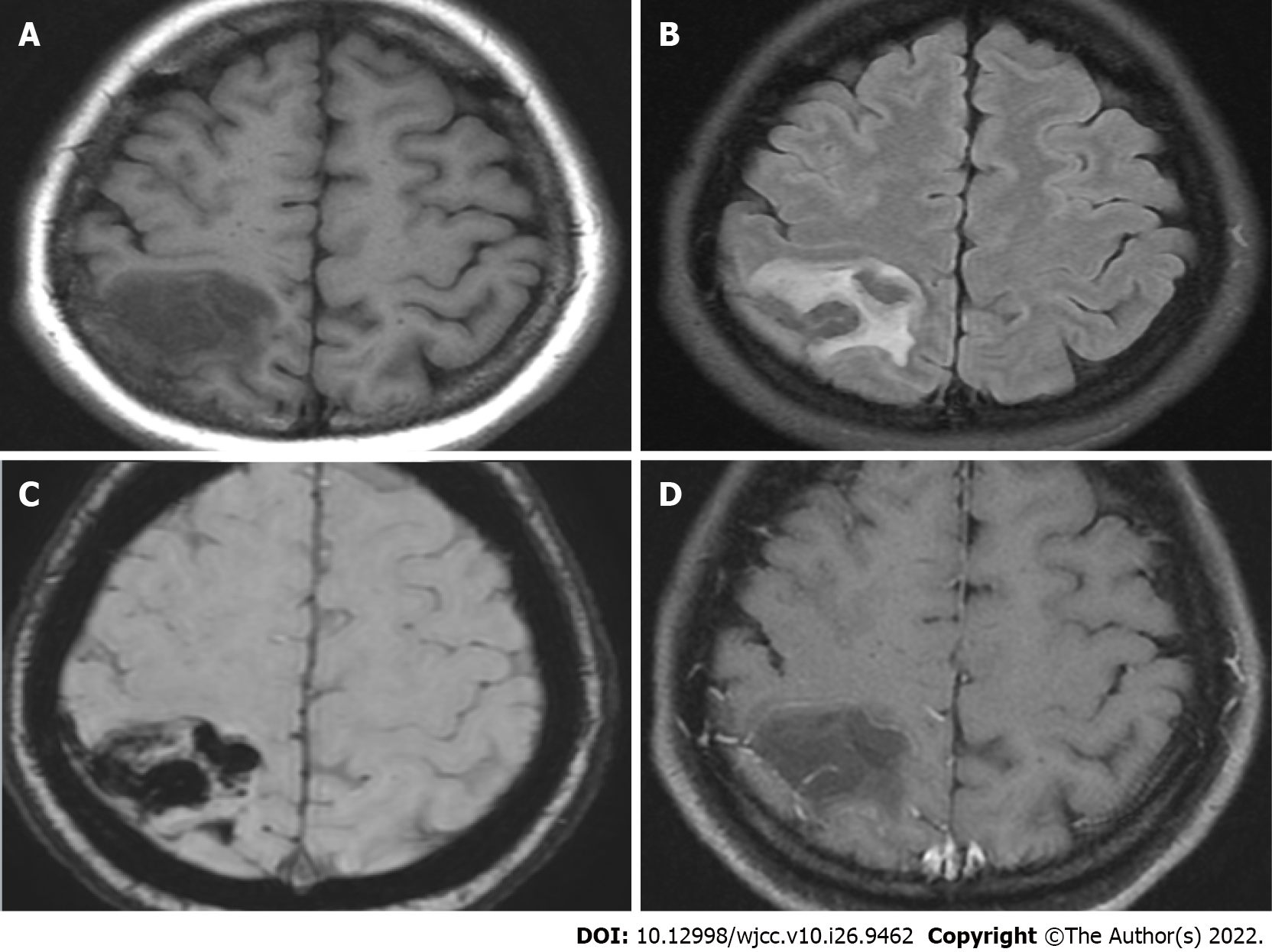
Figure 3 Brain magnetic resonance imaging at two hours after the initial brain computed tomography.
These series demonstrated hyperacute hemorrhage at the right parietal lobe. A: T1 image showing a hypointense lesion at the right parietal lobe; B: T2 FLAIR showing a hyperintense lesion; C: SWAN image showing hypointense “black dots” at the right parietal lobe; D: T1 with gadolinium enhancement did not enhance the lesion. FLAIR: Fluid-attenuated inversion recovery; SWAN: Susceptibility-weighted angiography.
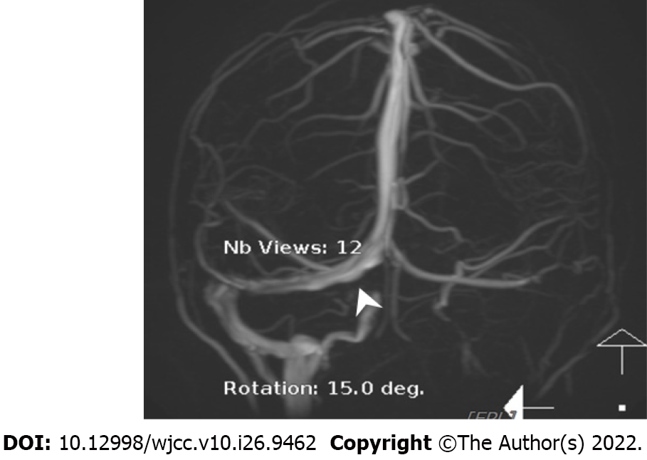
Figure 4 Magnetic resonance venography.
Arrowhead: An irregular contour of the right transverse sinus is noted.
Figure 5 Follow-up brain computed tomography on day 3 of hospitalization.
Arrowhead: Hyperdense acute hemorrhage at the right parietal lobe, in resolution. Asterisk: Hypodense perifocal edema around the acute hemorrhage, indicating the early phase of hematoma absorption.
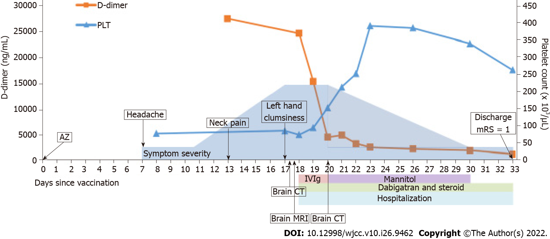
Figure 6 The clinical time course after vaccination with ChAdOx1 nCoV-19 vaccine (AZD1222).
AZ: AstraZeneca; ChAdOx1 nCoV-19 vaccine (AZD1222); PLT: Platelet; IVIG: Intravenous immunoglobulin; mRS: Modified Rankin scale; MRI: Magnetic resonance imaging.
- Citation: Jiang SK, Chen WL, Chien C, Pan CS, Tsai ST. Rapid progressive vaccine-induced immune thrombotic thrombocytopenia with cerebral venous thrombosis after ChAdOx1 nCoV-19 (AZD1222) vaccination: A case report. World J Clin Cases 2022; 10(26): 9462-9469
- URL: https://www.wjgnet.com/2307-8960/full/v10/i26/9462.htm
- DOI: https://dx.doi.org/10.12998/wjcc.v10.i26.9462


