Copyright
©The Author(s) 2022.
World J Clin Cases. Sep 16, 2022; 10(26): 9454-9461
Published online Sep 16, 2022. doi: 10.12998/wjcc.v10.i26.9454
Published online Sep 16, 2022. doi: 10.12998/wjcc.v10.i26.9454
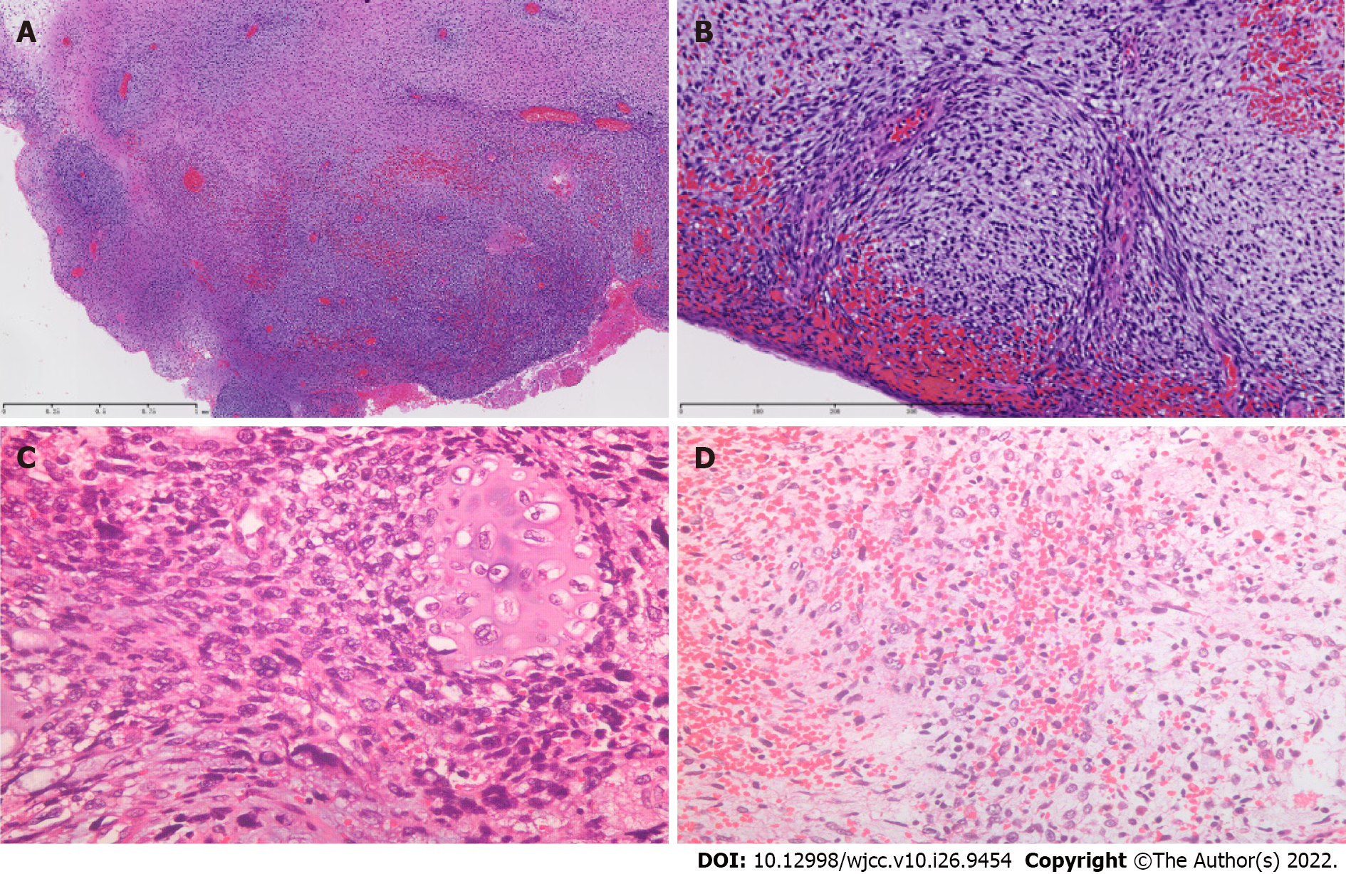
Figure 1 Pathologic examination of cervical embryonal rhabdomyosarcoma.
A: Lower power view of the tumor cells; B: Higher power view of the tumor cells; C: Cartilage component (200 ×); D: Primitive stellate cells (200 ×).
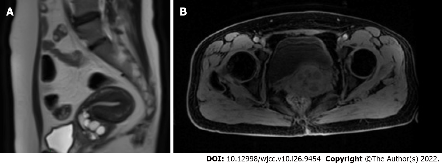
Figure 2 Magnetic resonance imaging of the pelvis before and after neoadjuvant chemotherapy.
A: The initial cervical mass size was about 5 cm; B: After neoadjuvant chemotherapy, the tumor size was reduced to 3 cm.
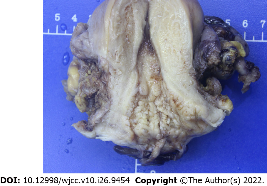
Figure 3
Gross appearance of the resected cervical tumor.
The neoplasm infiltrates the cervical walls and grows in a polypoid fashion in the posterior cervical lip.
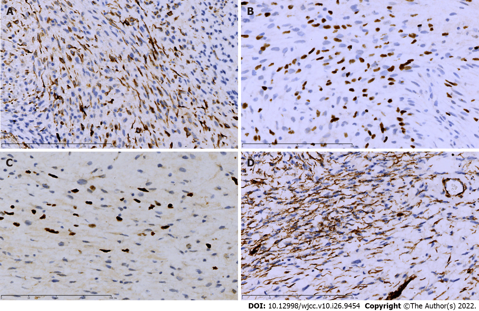
Figure 4 Immunohistochemistry showed in the tumor cells cytoplasmic positivity for Desmin, MyoD1, Myogenin, and WT-1.
A: Desmin; B: MyoD1; C: Myogenin; D: WT1.
- Citation: Xu LJ, Cai J, Huang BX, Dong WH. Locally advanced cervical rhabdomyosarcoma in adults: A case report. World J Clin Cases 2022; 10(26): 9454-9461
- URL: https://www.wjgnet.com/2307-8960/full/v10/i26/9454.htm
- DOI: https://dx.doi.org/10.12998/wjcc.v10.i26.9454


