Copyright
©The Author(s) 2022.
World J Clin Cases. Sep 16, 2022; 10(26): 9447-9453
Published online Sep 16, 2022. doi: 10.12998/wjcc.v10.i26.9447
Published online Sep 16, 2022. doi: 10.12998/wjcc.v10.i26.9447
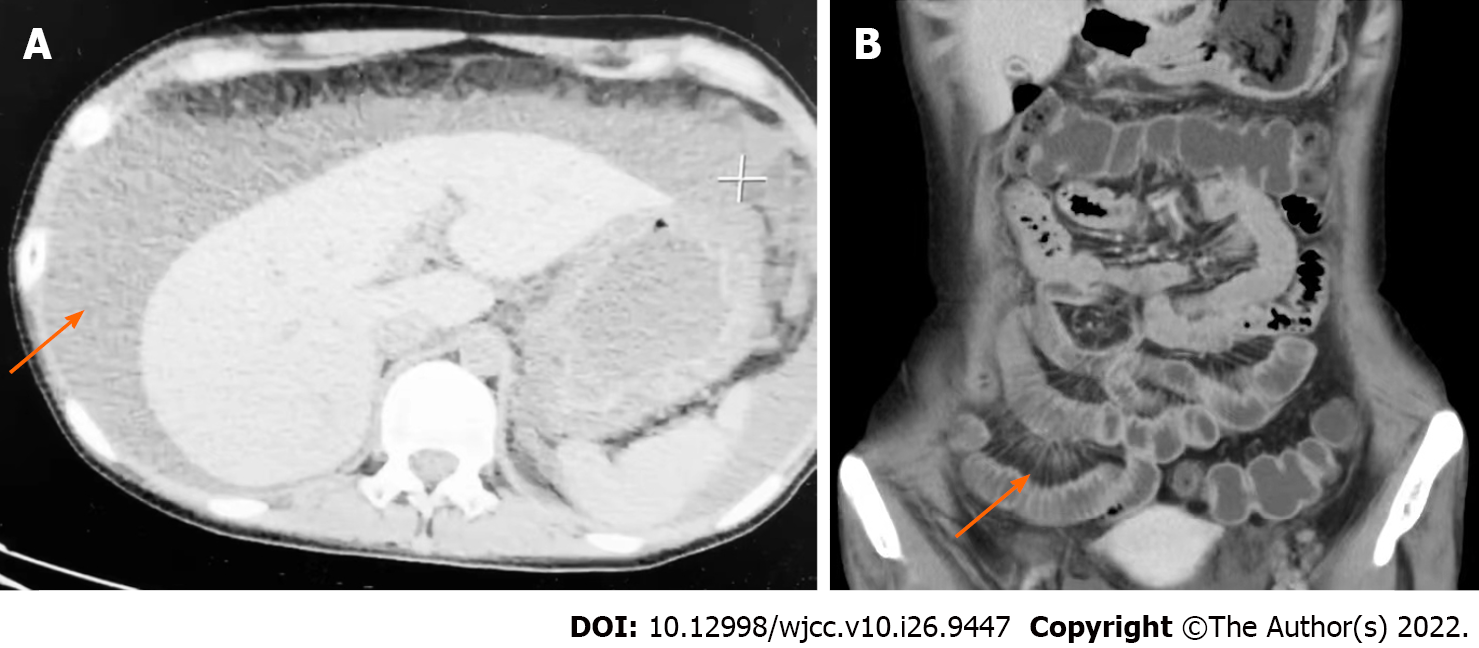
Figure 1 Computed tomography scan.
A: Abdominal computed tomography scan revealed massive ascites (orange arrow); B: Small bowel enhanced computed tomography revealed that the number of mesenteric vessels was increased. Mesenteric vessels were engorged and exhibited a “comb sign” (orange arrow) appearance.
- Citation: Wang JD, Yang YF, Zhang XF, Huang J. Systemic lupus erythematosus presenting with progressive massive ascites and CA-125 elevation indicating Tjalma syndrome? A case report. World J Clin Cases 2022; 10(26): 9447-9453
- URL: https://www.wjgnet.com/2307-8960/full/v10/i26/9447.htm
- DOI: https://dx.doi.org/10.12998/wjcc.v10.i26.9447


