Copyright
©The Author(s) 2022.
World J Clin Cases. Sep 16, 2022; 10(26): 9404-9410
Published online Sep 16, 2022. doi: 10.12998/wjcc.v10.i26.9404
Published online Sep 16, 2022. doi: 10.12998/wjcc.v10.i26.9404
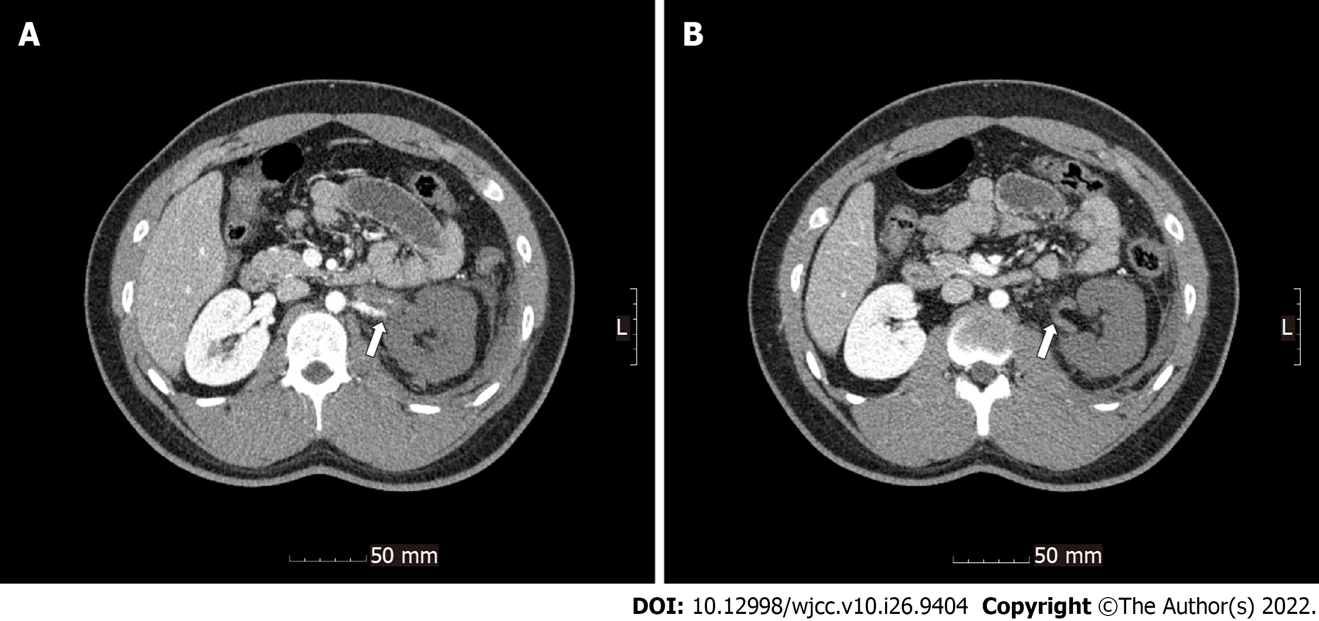
Figure 1 Renal infarction seen in the initial computed tomography scan.
A: Occlusion of the left renal artery with global infarction of the left kidney; B: Left renal artery without blood flow.
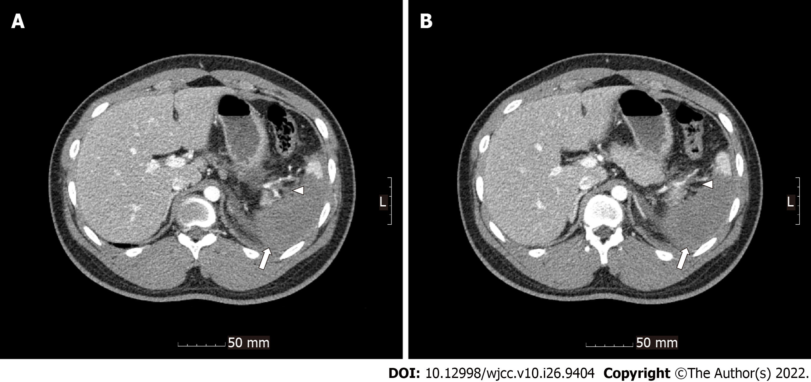
Figure 2 Splenic infarction seen in the initial computed tomography scan.
A and B: Multiple occlusions of the branches of the splenic artery (arrowhead) with infarction of the central portion of the spleen (arrow).
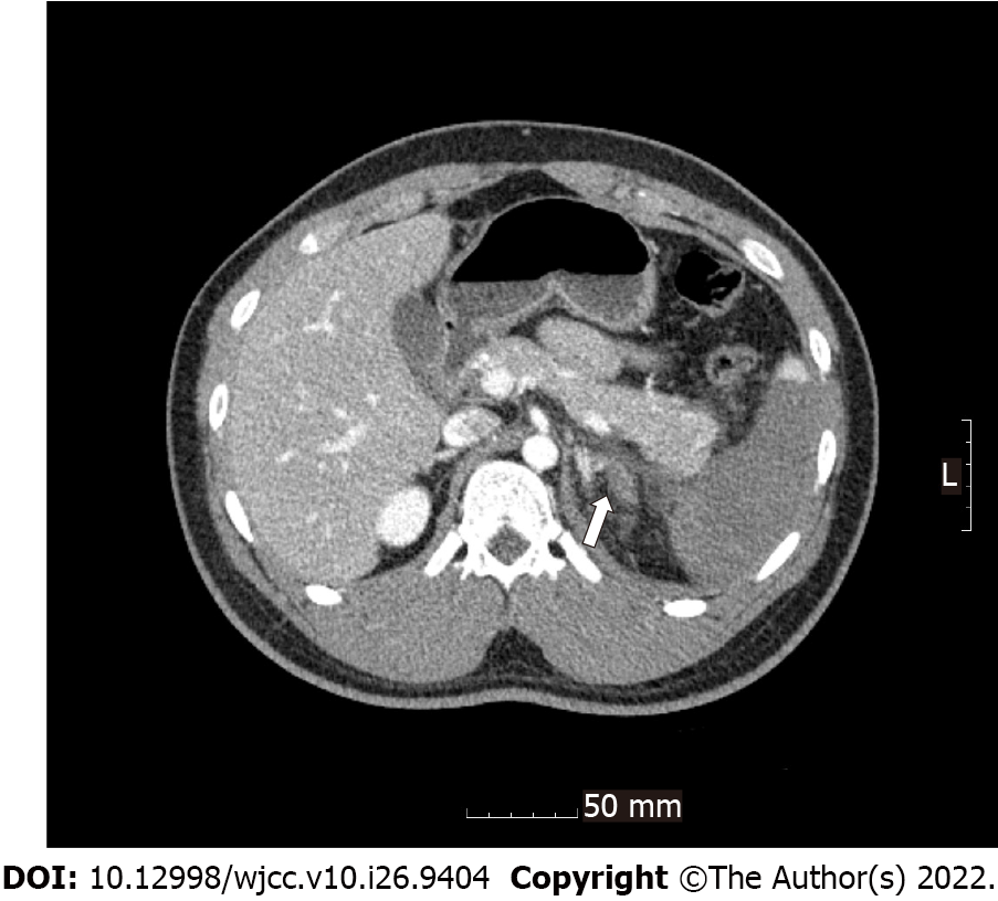
Figure 3
Left adrenal hematoma seen in the initial computed tomography scan.
- Citation: Lee NA, Jeong ES, Jang HS, Park YC, Kang JH, Kim JC, Jo YG. Antiphospholipid syndrome with renal and splenic infarction after blunt trauma: A case report. World J Clin Cases 2022; 10(26): 9404-9410
- URL: https://www.wjgnet.com/2307-8960/full/v10/i26/9404.htm
- DOI: https://dx.doi.org/10.12998/wjcc.v10.i26.9404


