Copyright
©The Author(s) 2022.
World J Clin Cases. Sep 16, 2022; 10(26): 9354-9360
Published online Sep 16, 2022. doi: 10.12998/wjcc.v10.i26.9354
Published online Sep 16, 2022. doi: 10.12998/wjcc.v10.i26.9354
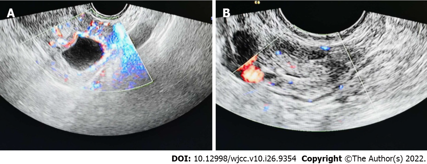
Figure 1 Heterogeneous echo of the uterine cavity.
A: The left ovary is a cystic echo with circumferential blood flow; B: In the hypoechoic area adjacent to the left adnexa, blood flow signals can be found in the periphery and in it.
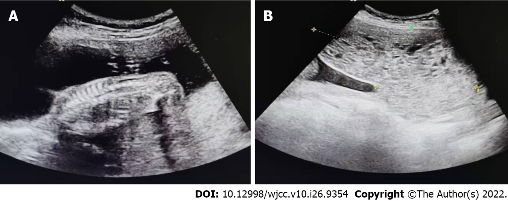
Figure 2 Partial hydatidiform mole.
A: Middle pregnancy, single live fetus; B: The anterior and inferior part of the fetal sac extends to the inner orifice of the cervix to see a heterogeneous echo area, showing a honeycomb shape, which is not clearly separated from the left placenta.
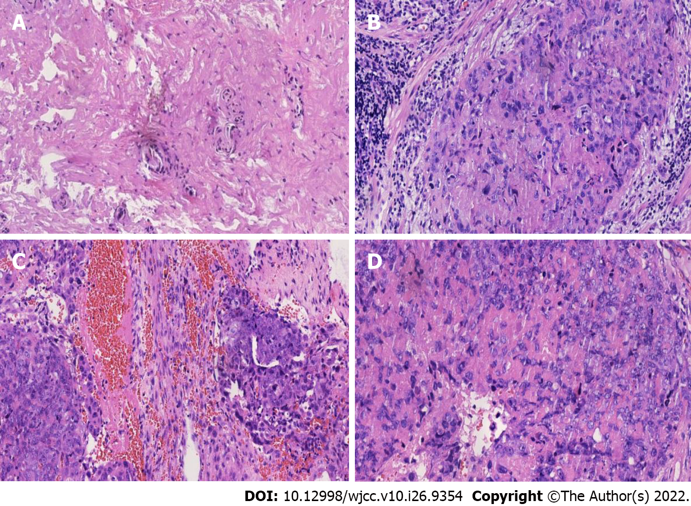
Figure 3 The postoperative pathological results.
A and B: Under the microscope, a small number of trophoblast epithelial cells were arranged in flakes, 2015; C: The tumor cells of epithelioid trophoblastic tumor are similar to chorionic intermediate trophoblast, 2020; D: Microscopy showing mononuclear intermediate trophoblastic cells, increased mitotic activity, and destructive invasion of the left ovary, 2020.
- Citation: Wang YN, Dong Y, Wang L, Chen YH, Hu HY, Guo J, Sun L. Special epithelioid trophoblastic tumor: A case report. World J Clin Cases 2022; 10(26): 9354-9360
- URL: https://www.wjgnet.com/2307-8960/full/v10/i26/9354.htm
- DOI: https://dx.doi.org/10.12998/wjcc.v10.i26.9354


