Copyright
©The Author(s) 2022.
World J Clin Cases. Sep 16, 2022; 10(26): 9348-9353
Published online Sep 16, 2022. doi: 10.12998/wjcc.v10.i26.9348
Published online Sep 16, 2022. doi: 10.12998/wjcc.v10.i26.9348
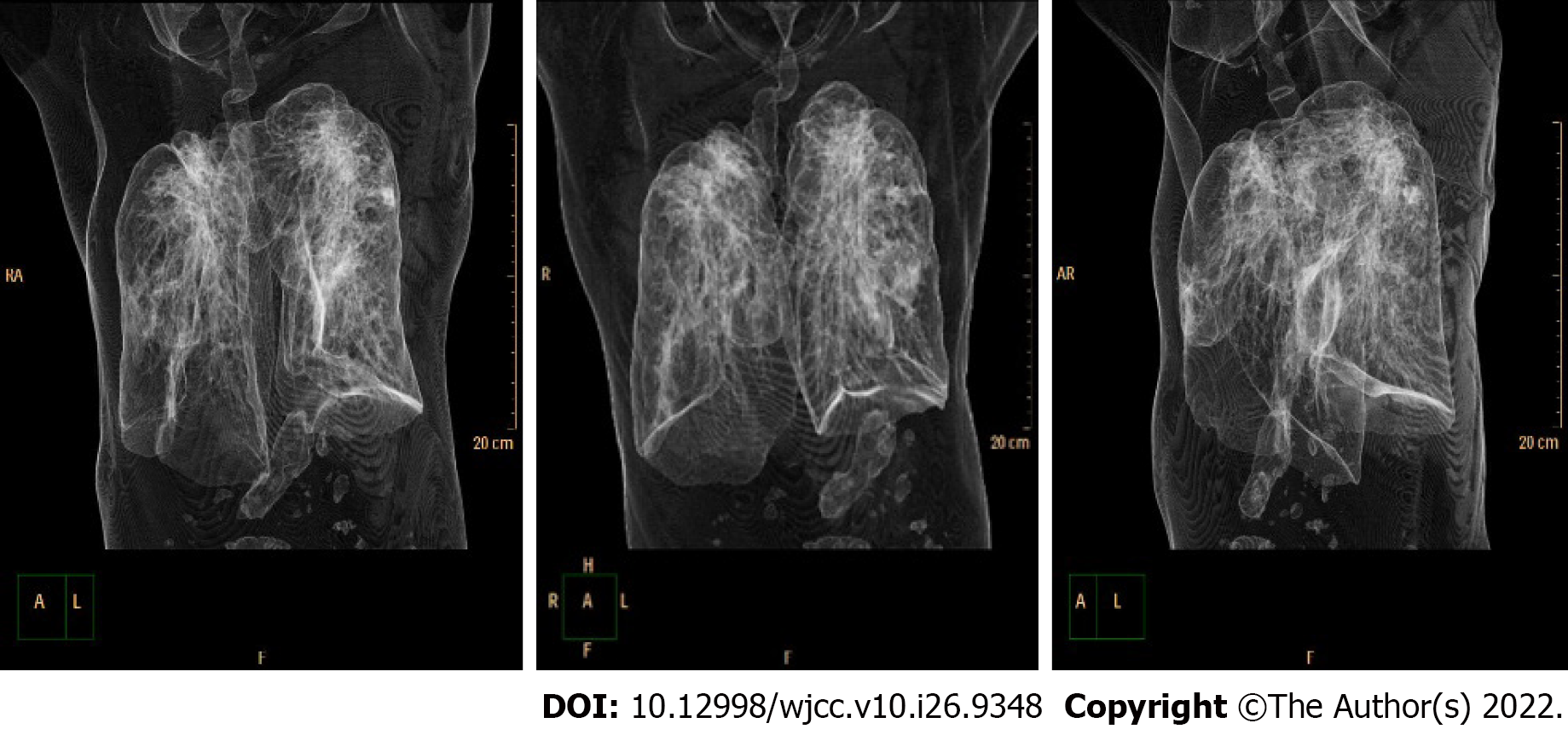
Figure 1
3D computed tomography scan image of trachea and lung.
Figure 2
3D computed tomography reconstruction of the trachea.
Figure 3 Image under bronchoscopy.
The blue arrow shows the site of distortion of the upper airway, the fiberoptic bronchoscopy figures are more intuitive.
- Citation: Zhou JW, Wang CG, Chen G, Zhou YF, Ding JF, Zhang JW. Unexpected difficult airway due to severe upper tracheal distortion: A case report. World J Clin Cases 2022; 10(26): 9348-9353
- URL: https://www.wjgnet.com/2307-8960/full/v10/i26/9348.htm
- DOI: https://dx.doi.org/10.12998/wjcc.v10.i26.9348


