Copyright
©The Author(s) 2022.
World J Clin Cases. Sep 16, 2022; 10(26): 9340-9347
Published online Sep 16, 2022. doi: 10.12998/wjcc.v10.i26.9340
Published online Sep 16, 2022. doi: 10.12998/wjcc.v10.i26.9340
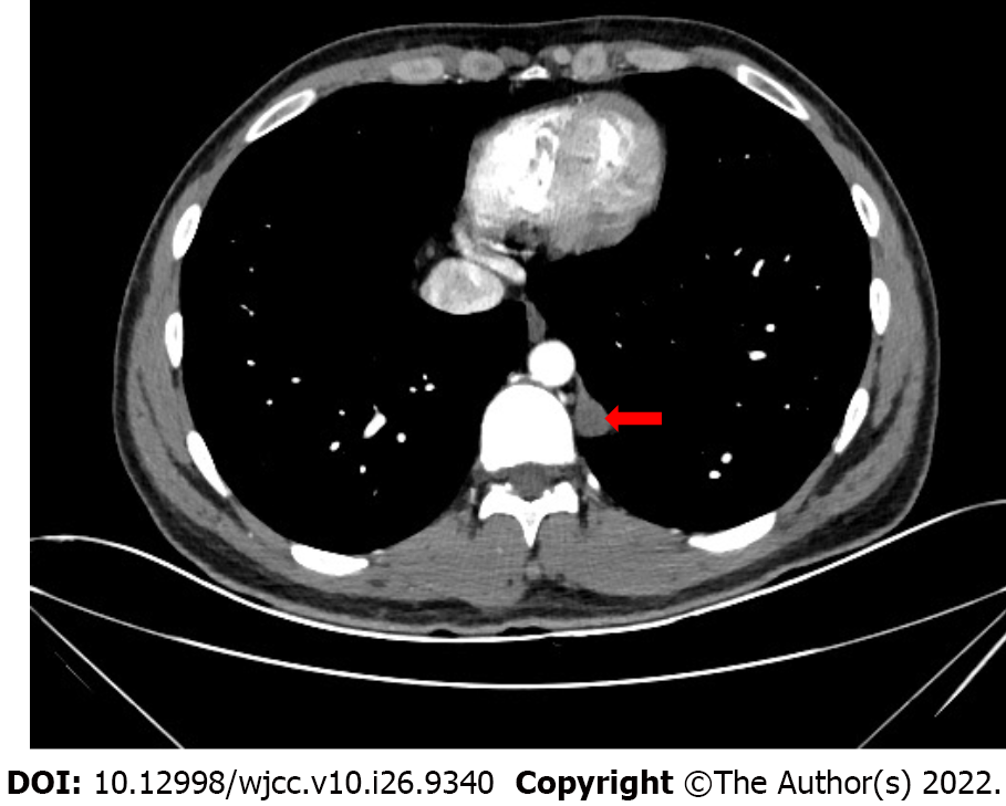
Figure 1 Chest computed tomography: Posterior mediastinal tumor measuring 1.
2 cm × 1.4 cm × 3.3 cm in size. The tumor consists of some cystic areas and shows slight enhancement in the arterial phase.
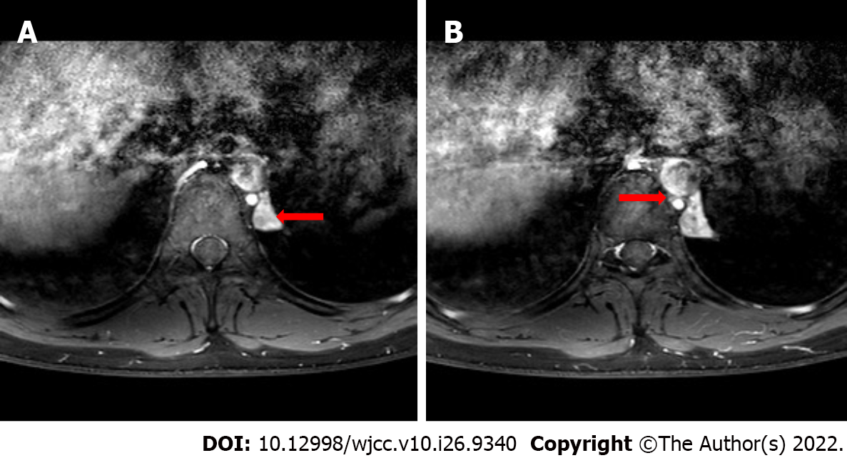
Figure 2 Chest magnetic resonance imaging.
A: The tumor shows heterogeneous enhancement after an enhanced scan (red arrow); B: The supplying vessel (red arrow) can be seen between the hemiazygos vein and the descending aorta.
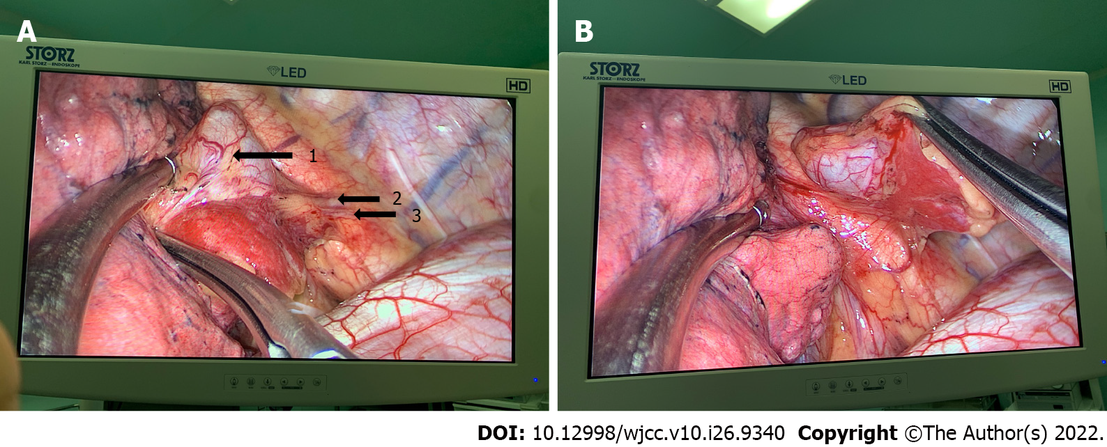
Figure 3 The tumor in the thoracoscopy.
A: The pyramidal tumor with two blood vessels can be seen in the posterior mediastinum (black arrow 1: Tumor; 2: Draining vein; 3: Supplying artery); B: The tumor has its own pleural covering and is isolated from the lung.
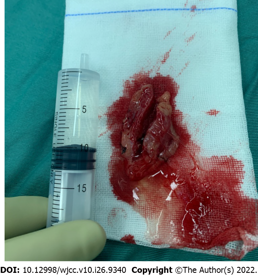
Figure 4 Complete resection of the tumor: Yellowish liquid was visible after cutting it open.
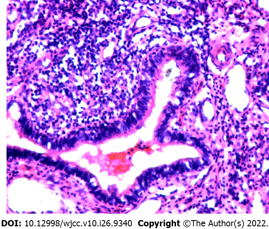
Figure 5 Pathology examination: Ciliated columnar epithelium, cartilage, and squamous cells lining the wall of the dilated, duct-like, cystic structure.
Obsolete hemorrhage and focal hyperplasia in the interstitial tissue are seen.
- Citation: Jin HJ, Yu Y, He W, Han Y. Posterior mediastinal extralobar pulmonary sequestration misdiagnosed as a neurogenic tumor: A case report. World J Clin Cases 2022; 10(26): 9340-9347
- URL: https://www.wjgnet.com/2307-8960/full/v10/i26/9340.htm
- DOI: https://dx.doi.org/10.12998/wjcc.v10.i26.9340


