Copyright
©The Author(s) 2022.
World J Clin Cases. Sep 16, 2022; 10(26): 9318-9322
Published online Sep 16, 2022. doi: 10.12998/wjcc.v10.i26.9318
Published online Sep 16, 2022. doi: 10.12998/wjcc.v10.i26.9318
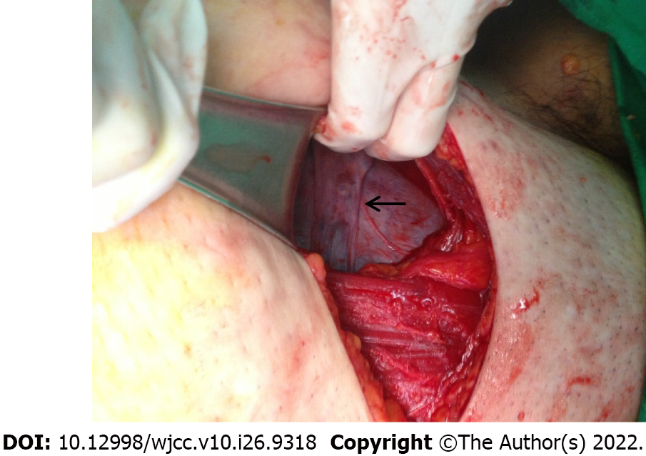
Figure 1
Thin rectangular-shaped tissue in front of the uterus (arrow).
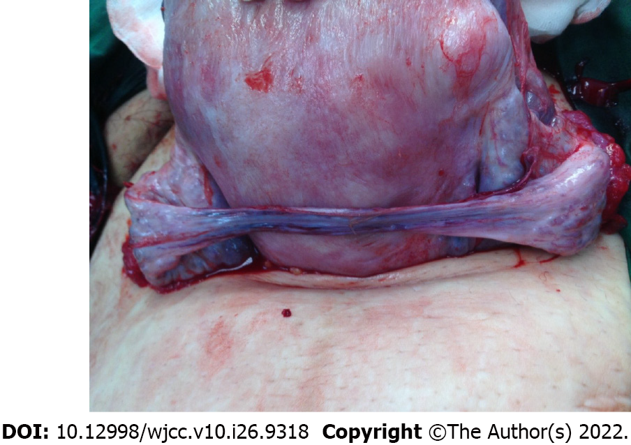
Figure 2
The tissue connected to both ovaries at the posterior view of the uterus after a cesarean section.
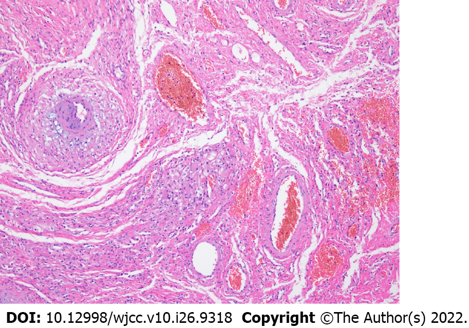
Figure 3
Well-vascularized loose stromal tissue with few follicles and scattered luteinized cells (x100).
- Citation: Choi MG, Kim JW, Kim YH, Kim AM, Kim TY, Ryu HK. Congenital ovarian anomaly manifesting as extra tissue connection between the two ovaries: A case report . World J Clin Cases 2022; 10(26): 9318-9322
- URL: https://www.wjgnet.com/2307-8960/full/v10/i26/9318.htm
- DOI: https://dx.doi.org/10.12998/wjcc.v10.i26.9318


