Copyright
©The Author(s) 2022.
World J Clin Cases. Sep 16, 2022; 10(26): 9310-9317
Published online Sep 16, 2022. doi: 10.12998/wjcc.v10.i26.9310
Published online Sep 16, 2022. doi: 10.12998/wjcc.v10.i26.9310
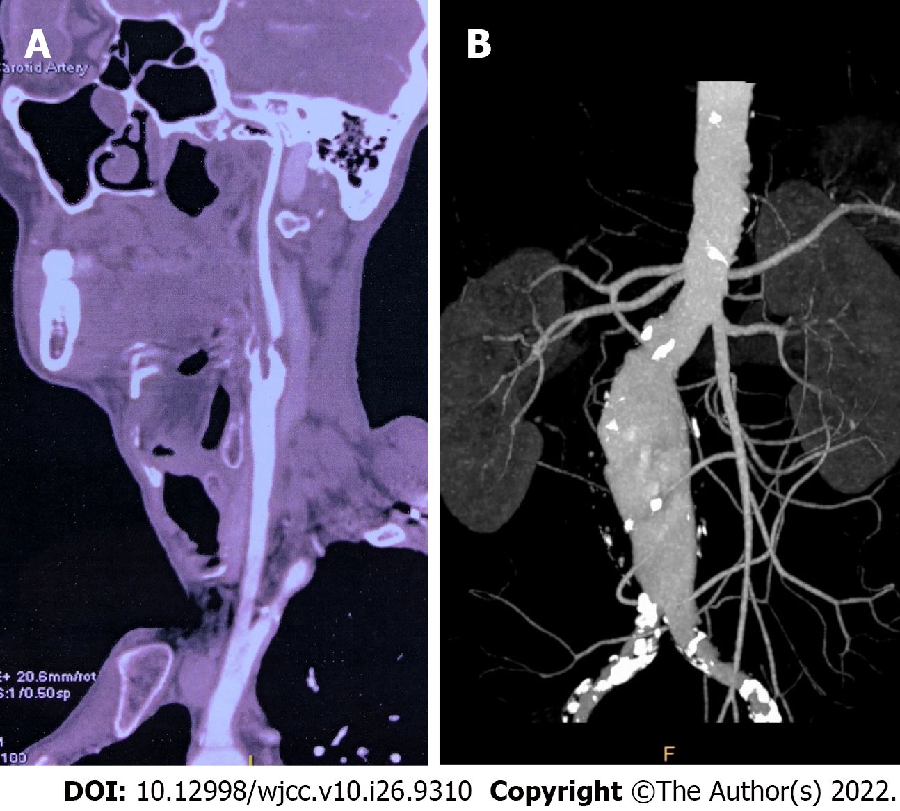
Figure 1 Pre-operative imaging examination.
A: Pre-operative computed tomography angioplasty (CTA) showed severe stenosis of right internal carotid artery; B: Aortic CTA showed infrarenal abdominal aortic aneurysm with maximum diameter of 53 mm.
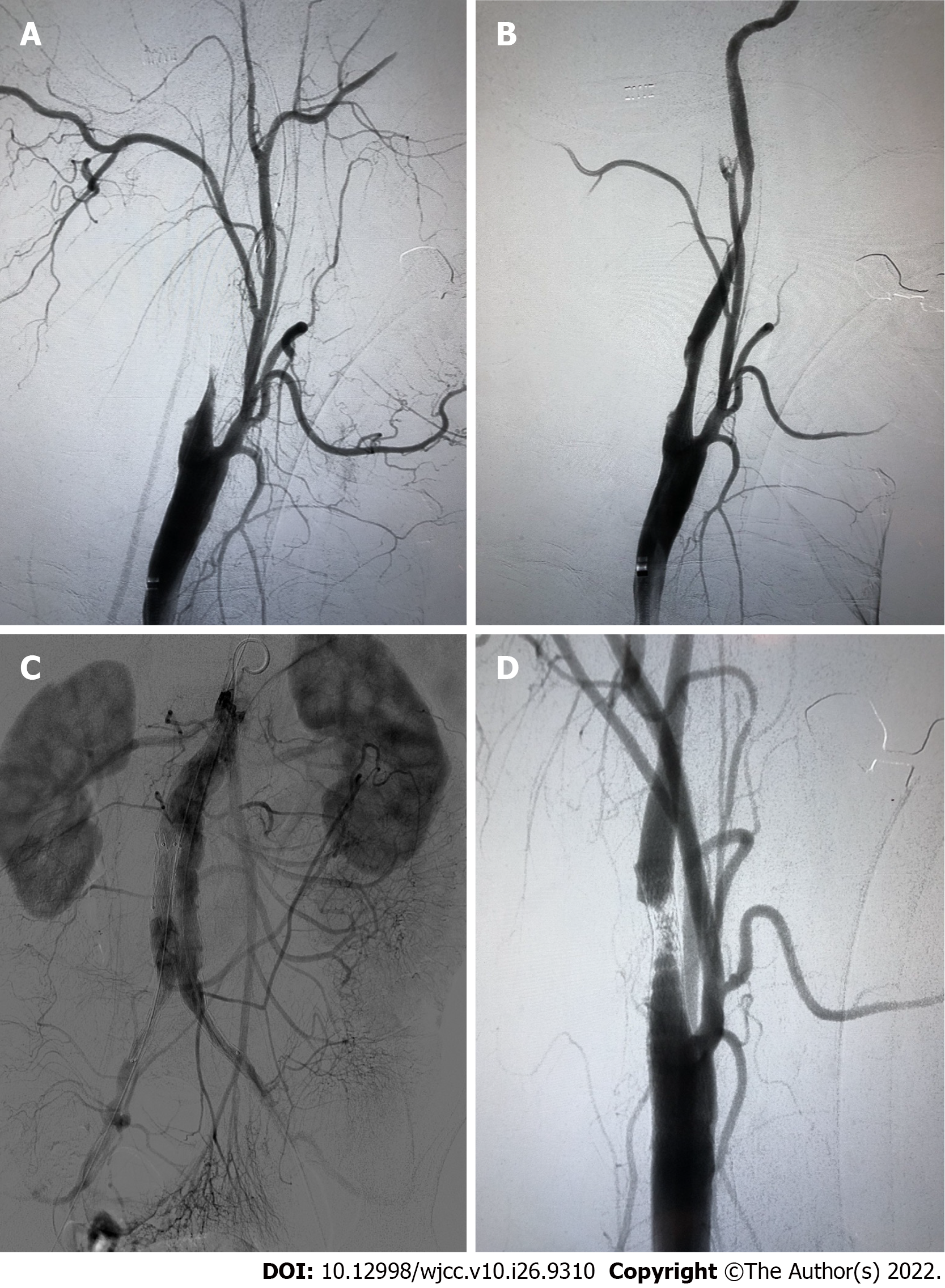
Figure 2 The interventional procedure.
A: The distal of the internal carotid artery (ICA) was obliterated; B: The ICA was patent with about 40% residual stenosis after the embolism protection device was retrieved; C: Angiography after endovascular aortic repair (EVAR); D: After EVAR, carotid angiography showed acute thrombus formation in the stent.
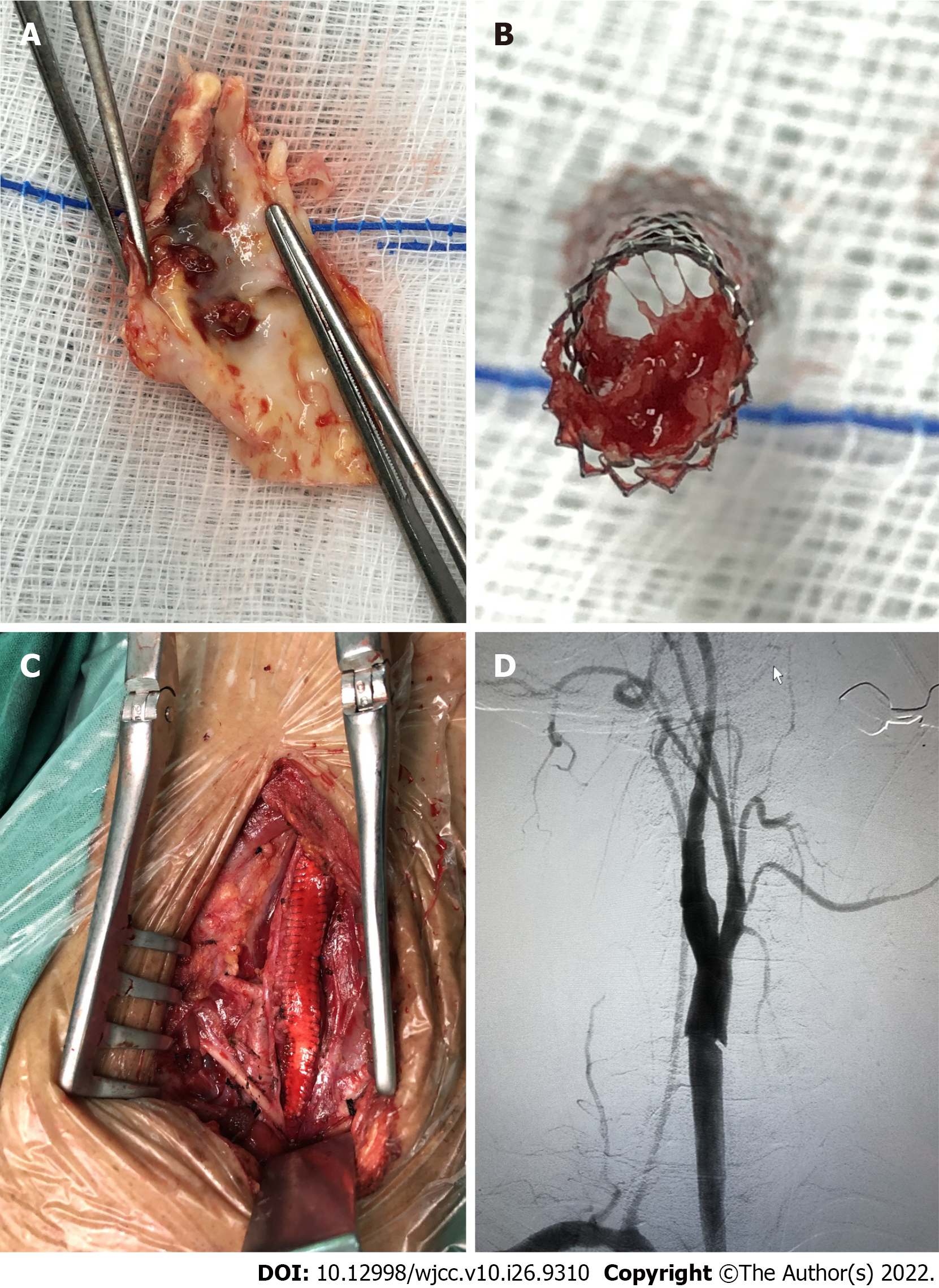
Figure 3 The carotid endarterectomy procedure.
A: Plaque rupture and thrombus formation; B: The stent was occluded by the plaque debris and thrombus; C: Patch angioplasty was carried out during carotid endarterectomy (CEA); D: Angiography after CEA showed the internal carotid artery was patent.
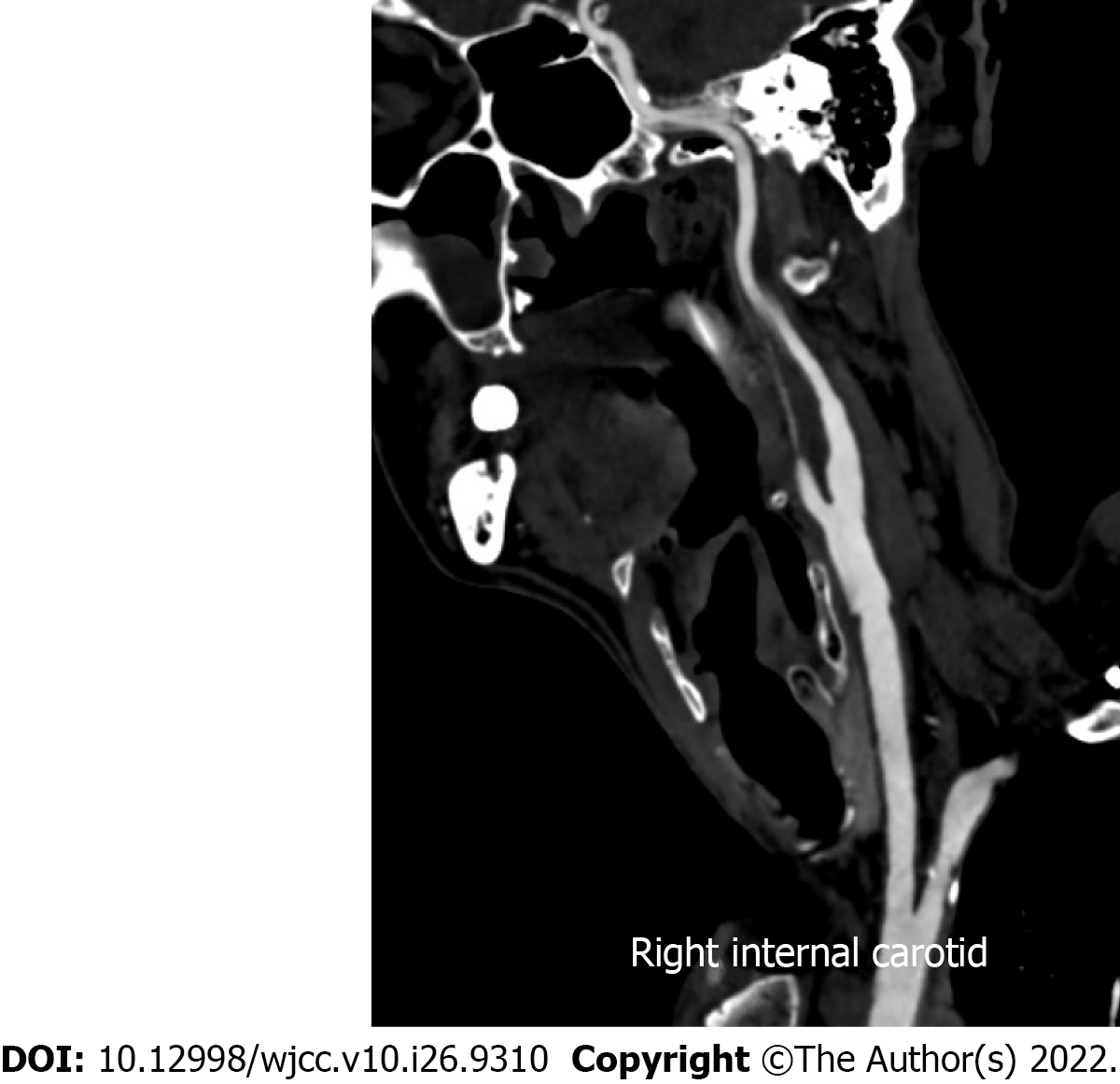
Figure 4
Computed tomography angioplasty after 1 yr showed the internal carotid artery was patent without restenosis.
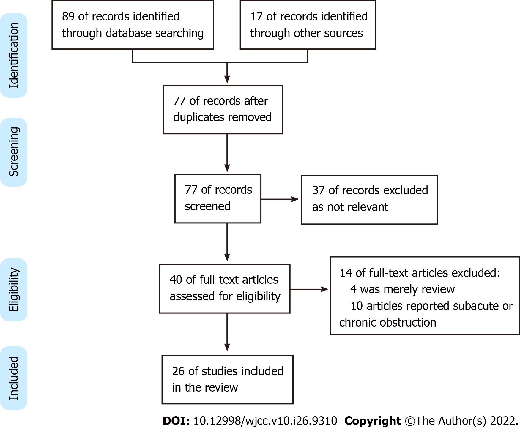
Figure 5
PRISMA flow chart for the study selection process.
- Citation: Zhang JB, Fan XQ, Chen J, Liu P, Ye ZD. Acute carotid stent thrombosis: A case report and literature review . World J Clin Cases 2022; 10(26): 9310-9317
- URL: https://www.wjgnet.com/2307-8960/full/v10/i26/9310.htm
- DOI: https://dx.doi.org/10.12998/wjcc.v10.i26.9310


