Copyright
©The Author(s) 2022.
World J Clin Cases. Sep 16, 2022; 10(26): 9276-9284
Published online Sep 16, 2022. doi: 10.12998/wjcc.v10.i26.9276
Published online Sep 16, 2022. doi: 10.12998/wjcc.v10.i26.9276
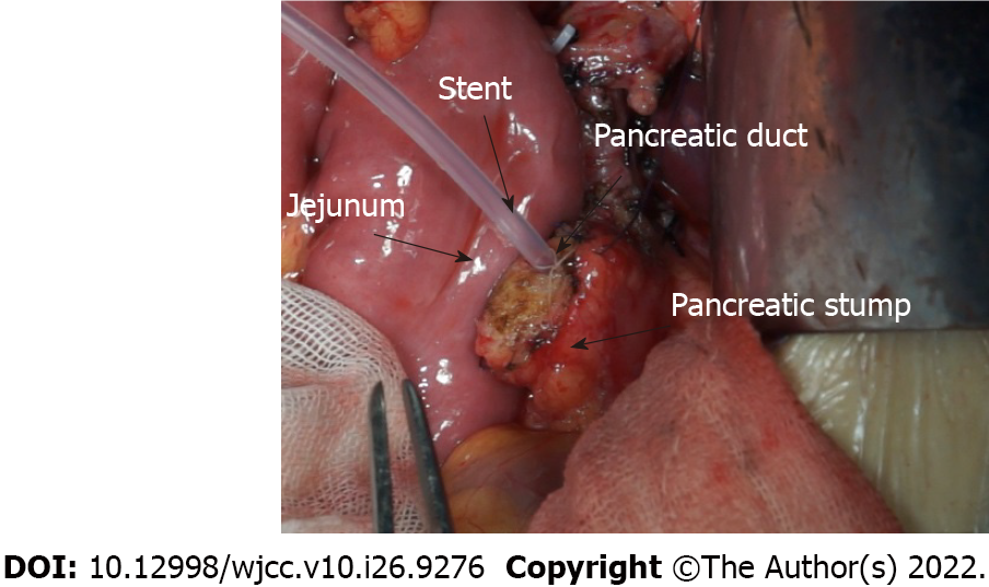
Figure 1 Pancreas and jejunal loop before anastomosis.
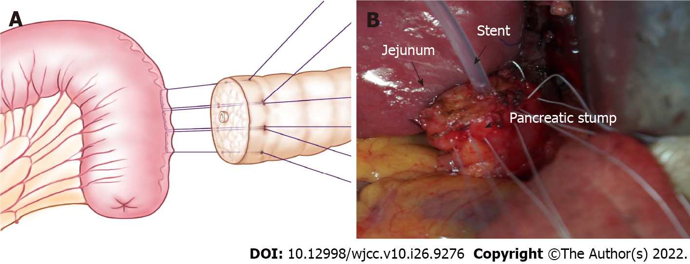
Figure 2 U-shaped sutures of a pancreaticojejunostomy.
A: One needle with 4-0 Gore thread is inserted through the anterior wall of the pancreas. The needle pierces through the pancreas and out from the rear wall with the same distance from the incisal margin. Then, suturing of the seromuscular layer of the jejunum loop continues along the direction of the intestine, which is approximately 1.5 cm beneath the anastomosis pore, and the distance is approximately 1/3 to 1/4 of the length of the pancreatic stump diameter. Finally, the needle pierces through the pancreas again from the rear wall to the anterior wall, ensuring that the needle point distance is approximately 1/3 to 1/4 of the pancreatic stump diameter; B: Photographic image of U-shape sutures.
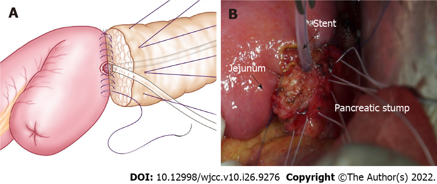
Figure 3 One-layer anastomosis of the pancreas and jejunum.
A: Continuous suturing with a 5-0 absorbable suture is used to complete the anastomosis between the seromuscular layer of the jejunum and the rear wall of the pancreas stump. A pancreatic duct-to-mucosa anastomosis is performed for the pancreatic duct; B: Photographic image of one-layer anastomosis.
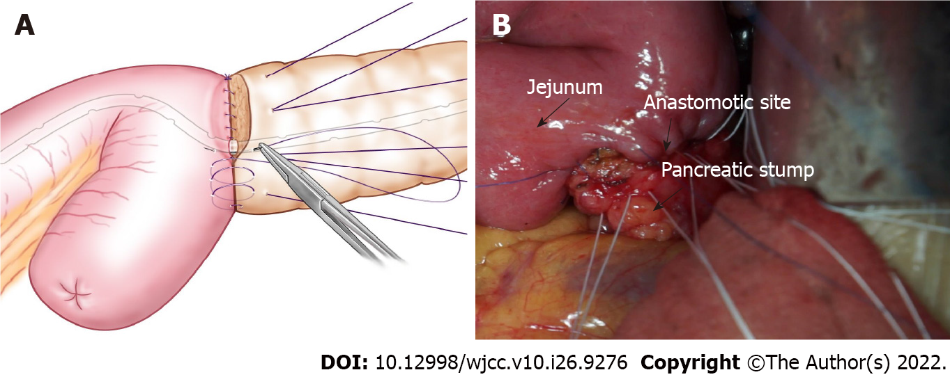
Figure 4 The anterior wall of one-layer anastomosis.
A: Suturing of the anterior wall of the anastomosis is continued similar to the rear wall to complete the one-layer match; B: Photographic image of the operation.
Figure 5 The rear wall of the pancreas was reinforced.
A: The 3 to 4-pin U-shaped sutures are ligated to hold the rear wall of the pancreas. The schema shows reinforcement of the rear wall of the pancreas; B: Photographic image shows the reinforcement of the rear wall of the pancreas; C: The schema of a longitudinal section after the rear wall of the pancreas is reinforced.
- Citation: Wei JP, Tai S, Su ZL. One-half layer pancreaticojejunostomy with the rear wall of the pancreas reinforced: A valuable anastomosis technique. World J Clin Cases 2022; 10(26): 9276-9284
- URL: https://www.wjgnet.com/2307-8960/full/v10/i26/9276.htm
- DOI: https://dx.doi.org/10.12998/wjcc.v10.i26.9276


