Copyright
©The Author(s) 2022.
World J Clin Cases. Sep 16, 2022; 10(26): 9264-9275
Published online Sep 16, 2022. doi: 10.12998/wjcc.v10.i26.9264
Published online Sep 16, 2022. doi: 10.12998/wjcc.v10.i26.9264
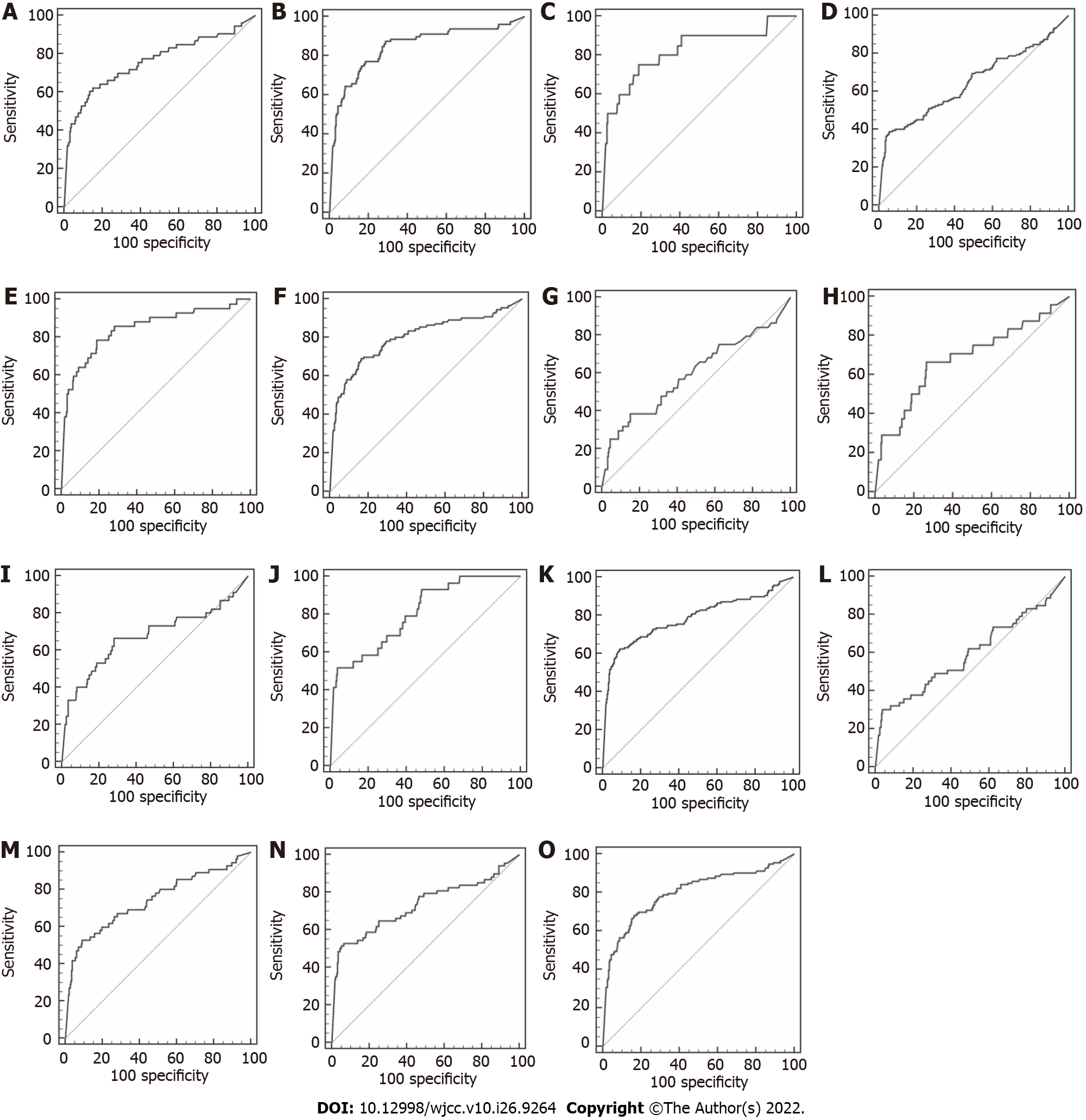
Figure 1 Receiver operating characteristic curves in different stratifications of primary liver cancer.
A-O: Represent primary liver cancer (PLC) of Child-Pugh A, PLC of Child-Pugh B, PLC of Child-Pugh C, PLC without cirrhosis, PLC with the compensated phase of cirrhosis, PLC with the decompensated phase of cirrhosis, small tumors, medium tumors, large tumors, massive tumors, diffuse tumors, PLC at Barcelona Clinic Liver Cancer (BCLC) stage A, PLC at BCLC stage B, PLC at BCLC stage C, and PLC at BCLC stage D, respectively. The contributions of related high-risk factors to stratified PLC were compared according to Child-Pugh score, clinical stage of concurrent liver cirrhosis, tumor size and BCLC stage, respectively.
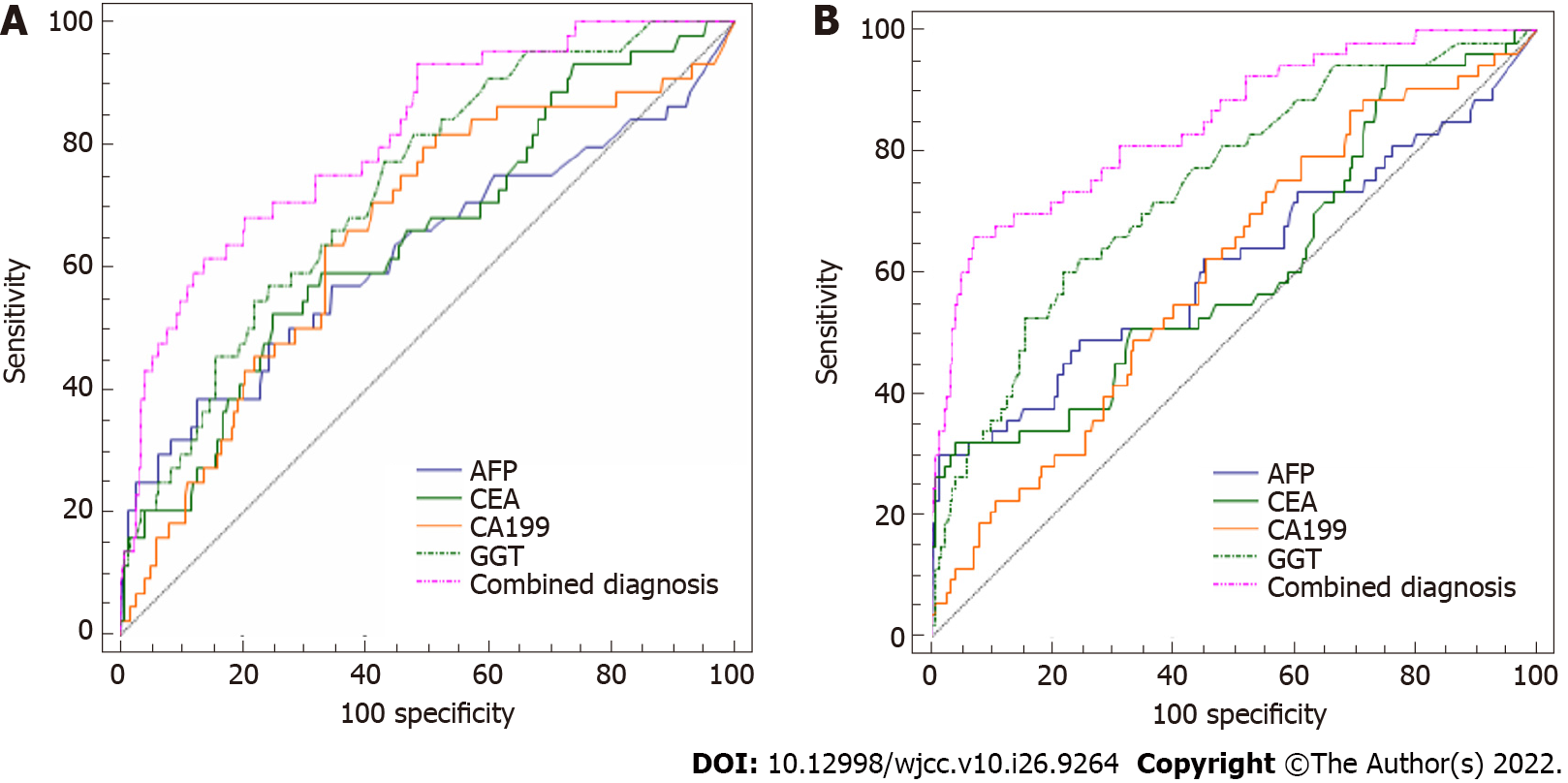
Figure 2 Area under the curve.
A-B: Represent small tumors and primary liver cancer (PLC) at Barcelona Clinic Liver Cancer (BCLC) stage A, respectively. Data above demonstrated that alpha-fetoprotein (AFP) had little diagnostic value in PLC patients with small tumors and at BCLC stage A. As shown, carcinoembryonic antigen, carbohydrate antigen 19-9, and gamma-glutamyl transferase improved the diagnostic value of AFP. AFP: Alpha-fetoprotein; BCLC: Barcelona Clinic Liver Cancer; CEA: Carcinoembryonic antigen; CA 19-9: Carbohydrate antigen 19-9; GGT: Gamma-glutamyl transferase.
- Citation: Jiao HB, Wang W, Guo MN, Su YL, Pang DQ, Wang BL, Shi J, Wu JH. Evaluation of high-risk factors and the diagnostic value of alpha-fetoprotein in the stratification of primary liver cancer. World J Clin Cases 2022; 10(26): 9264-9275
- URL: https://www.wjgnet.com/2307-8960/full/v10/i26/9264.htm
- DOI: https://dx.doi.org/10.12998/wjcc.v10.i26.9264


