Copyright
©The Author(s) 2022.
World J Clin Cases. Sep 6, 2022; 10(25): 9156-9161
Published online Sep 6, 2022. doi: 10.12998/wjcc.v10.i25.9156
Published online Sep 6, 2022. doi: 10.12998/wjcc.v10.i25.9156
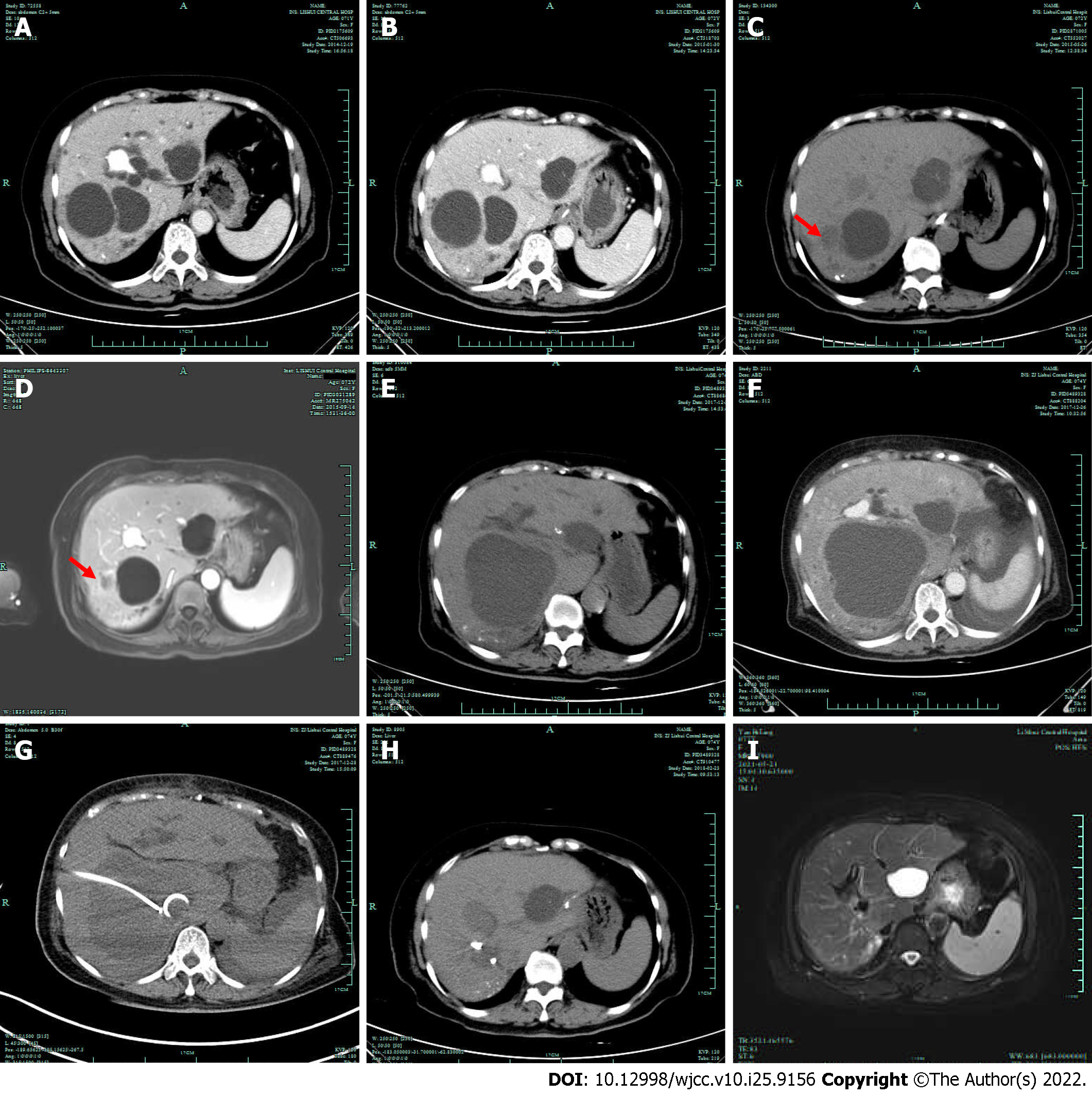
Figure 1 Imaging examinations.
A: Computed tomography (CT) revealed 3 Large cysts and some small cysts; B: Abdominal CT performed 2 mo postoperatively did not reveal signs of hepatic cyst infection; C and D: CT and magnetic resonance imaging (MRI) both showed that the size of the cyst on the lateral side of the right liver (arrow) was also significantly reduced, and the wall of the cyst was slightly thickened. Abdominal CT performed 5 mo postoperatively (C); abdominal MRI performed 9 mo postoperatively (D); E: Three years postoperatively, a plain CT scan revealed that the cyst on the lateral side of the right liver had disappeared and that the size of the cyst on the inner side of the right lobe had increased obviously; F: Enhanced CT scan revealed that the cyst had a thick wall and was surrounded by edema, and there was less pleural and peritoneal effusion; G: Percutaneous transhepatic drainage of the cyst under CT guidance; H: The infectious cyst disappeared 2 mo after percutaneous transhepatic drainage; I: There was no recurrence of cyst infection 4 years after drainage.
- Citation: Zhang K, Zhang HL, Guo JQ, Tu CY, Lv XL, Zhu JD. Repeated bacteremia and hepatic cyst infection lasting 3 years following pancreatoduodenectomy: A case report. World J Clin Cases 2022; 10(25): 9156-9161
- URL: https://www.wjgnet.com/2307-8960/full/v10/i25/9156.htm
- DOI: https://dx.doi.org/10.12998/wjcc.v10.i25.9156


