Copyright
©The Author(s) 2022.
World J Clin Cases. Sep 6, 2022; 10(25): 9142-9147
Published online Sep 6, 2022. doi: 10.12998/wjcc.v10.i25.9142
Published online Sep 6, 2022. doi: 10.12998/wjcc.v10.i25.9142
Figure 1 Cervical spine magnetic resonance imaging findings at admission.
A: T2 sagittal image. Cord signal changes at C4-C6 levels were observed; B: T2 axial image at C4 level. Cord signal changes at C4 level are shown; C: T2 axial image at C5 level. Cord signal changes at C5 level and possible hematoma were observed; D: T2 axial image at C6 level. Cord signal changes at C6 level are shown.
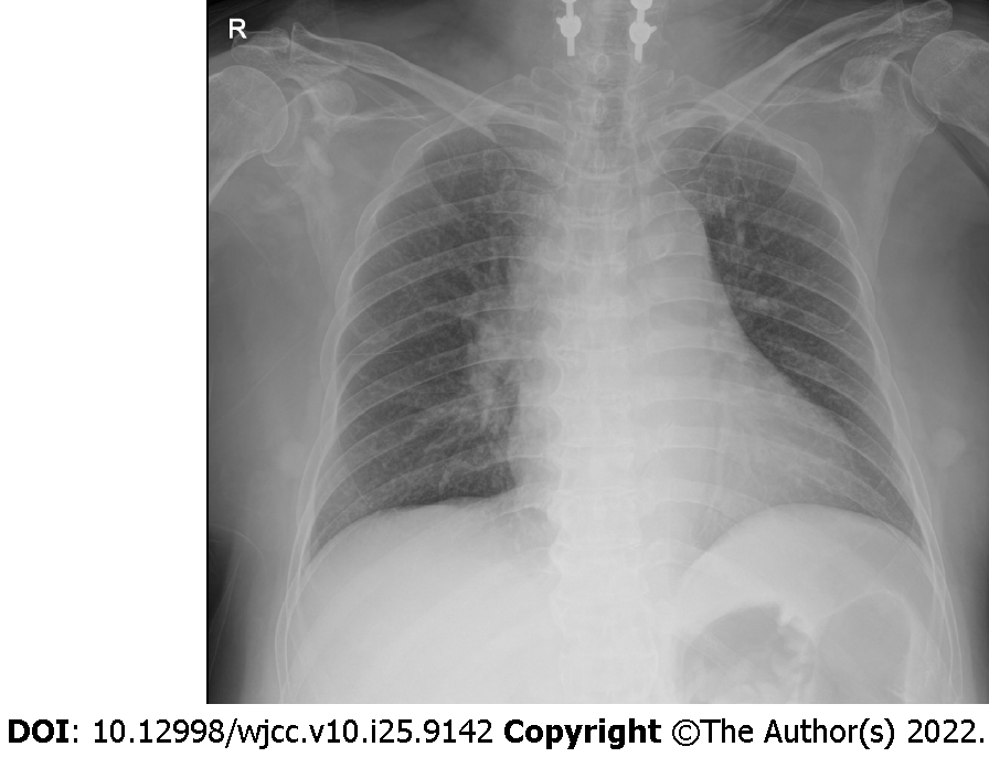
Figure 2 Chest X-ray at the first hypotensive event.
No specific findings were revealed at the event.
- Citation: Lee JY, Lee HS, Park SB, Lee KH. Tamsulosin-induced life-threatening hypotension in a patient with spinal cord injury: A case report. World J Clin Cases 2022; 10(25): 9142-9147
- URL: https://www.wjgnet.com/2307-8960/full/v10/i25/9142.htm
- DOI: https://dx.doi.org/10.12998/wjcc.v10.i25.9142


