Copyright
©The Author(s) 2022.
World J Clin Cases. Sep 6, 2022; 10(25): 9078-9086
Published online Sep 6, 2022. doi: 10.12998/wjcc.v10.i25.9078
Published online Sep 6, 2022. doi: 10.12998/wjcc.v10.i25.9078
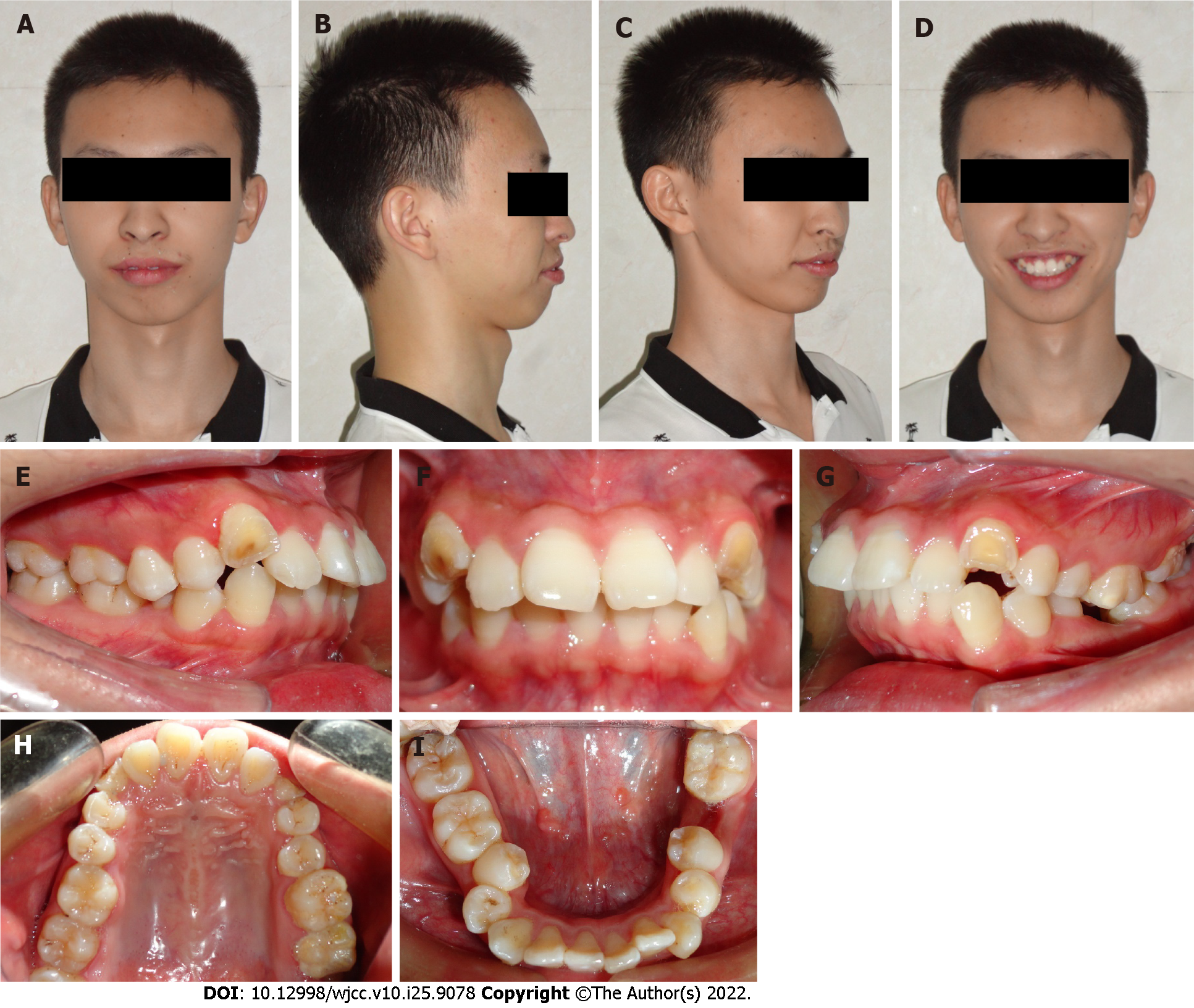
Figure 1 Pretreatment photographs.
A-D: Facial photographs; E-I: Intraoral photographs.
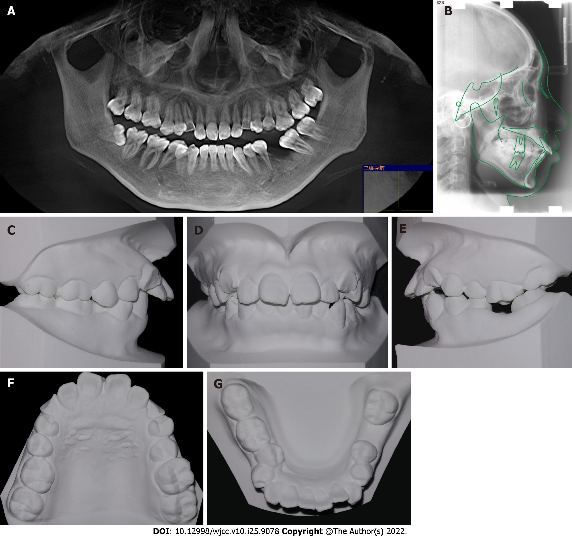
Figure 2 Pretreatment images.
A: Pretreatment panoramic radiograph; B: Pretreatment lateral head film and tracing; C-G: Pretreatment dental casts.
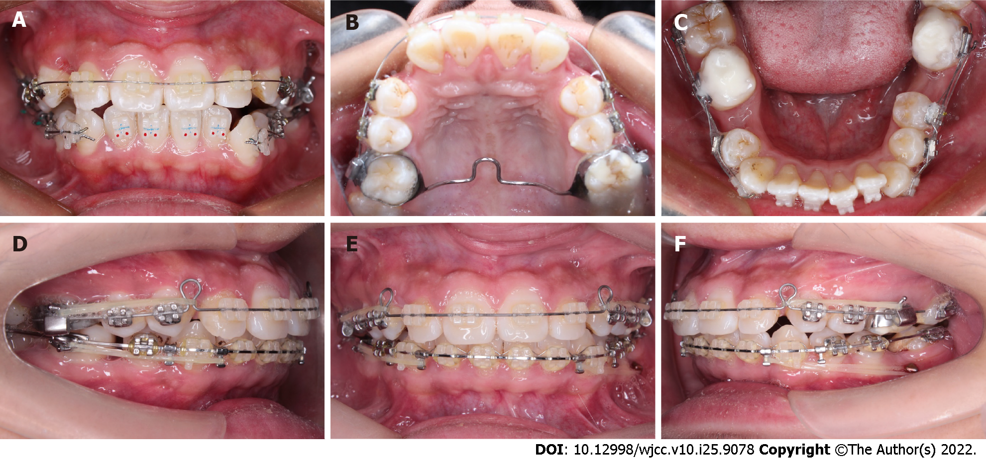
Figure 3 Progress intraoral photographs.
A-C: Leveling and aligning; D-F: Closing the extraction spaces.
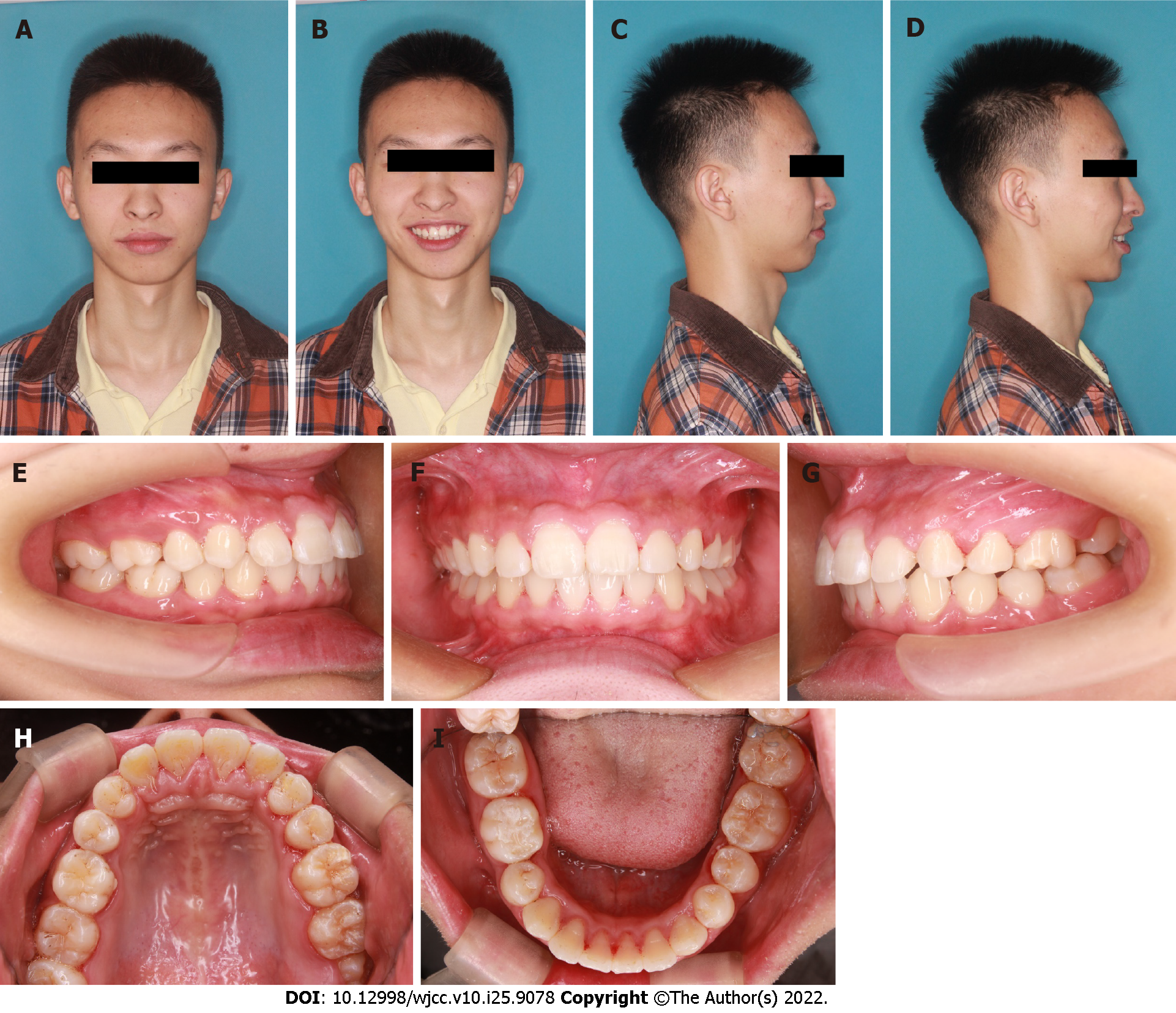
Figure 4 Posttreatment photographs.
A-D: Facial photographs ; E-I: Intraoral photographs.
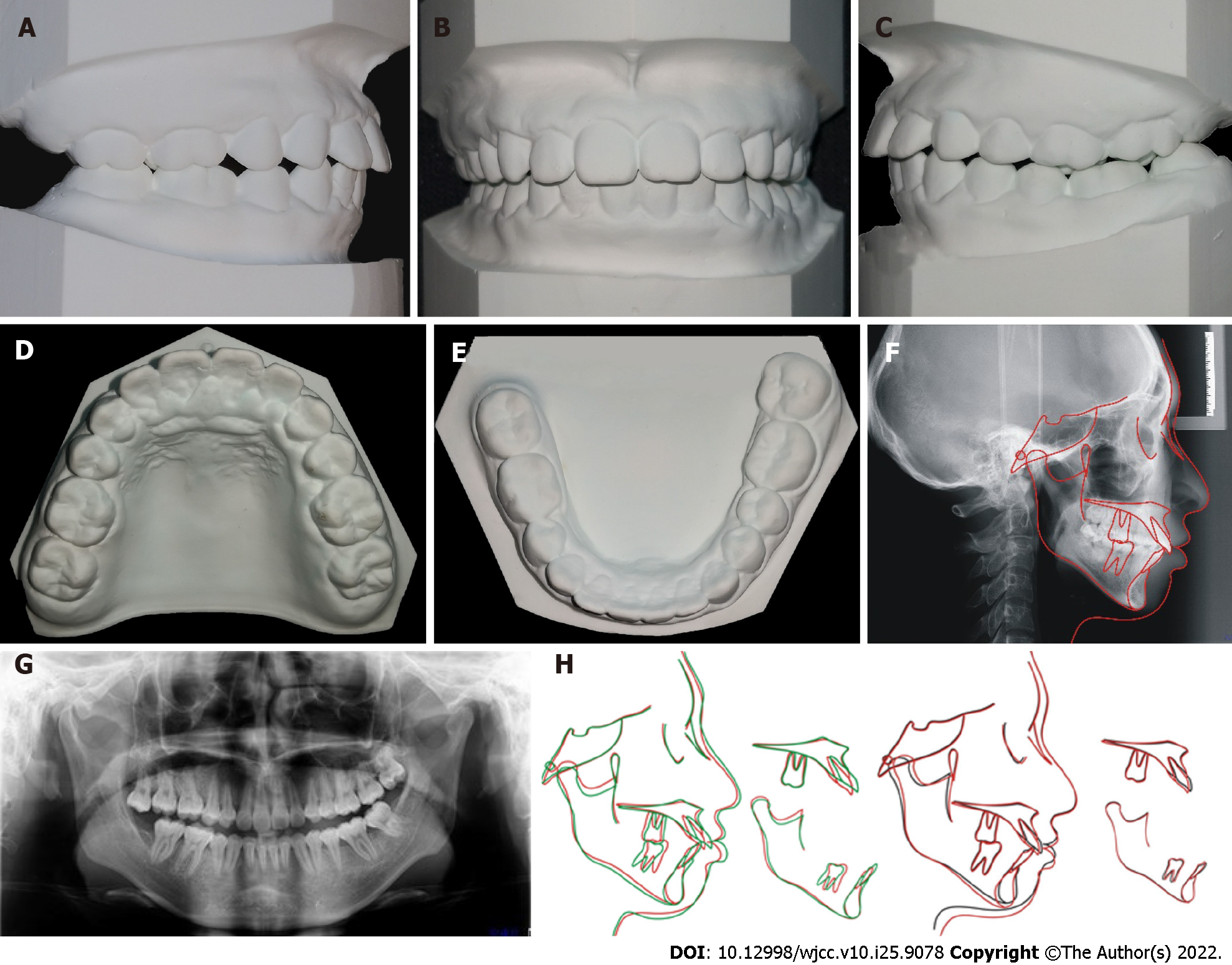
Figure 5 Posttreatment.
A-E: Posttreatment dental casts; F: Posttreatment lateral headfilm and tracing; G: Posttreatment panoramic radiograph; H: Superimposed tracings.
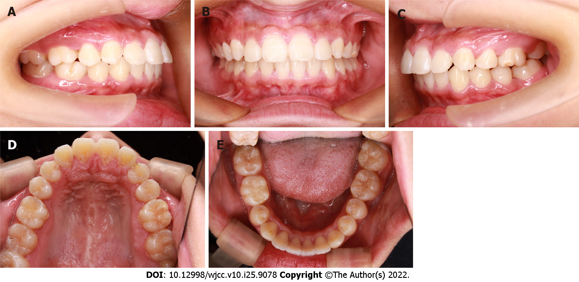
Figure 6 Photographs obtained at 3 years after the end of treatment.
A-E: Intraoral photographs.
- Citation: Li FF, Li M, Li M, Yang X. Modified orthodontic treatment of substitution of canines by first premolars: A case report. World J Clin Cases 2022; 10(25): 9078-9086
- URL: https://www.wjgnet.com/2307-8960/full/v10/i25/9078.htm
- DOI: https://dx.doi.org/10.12998/wjcc.v10.i25.9078


