Copyright
©The Author(s) 2022.
World J Clin Cases. Sep 6, 2022; 10(25): 8980-8989
Published online Sep 6, 2022. doi: 10.12998/wjcc.v10.i25.8980
Published online Sep 6, 2022. doi: 10.12998/wjcc.v10.i25.8980
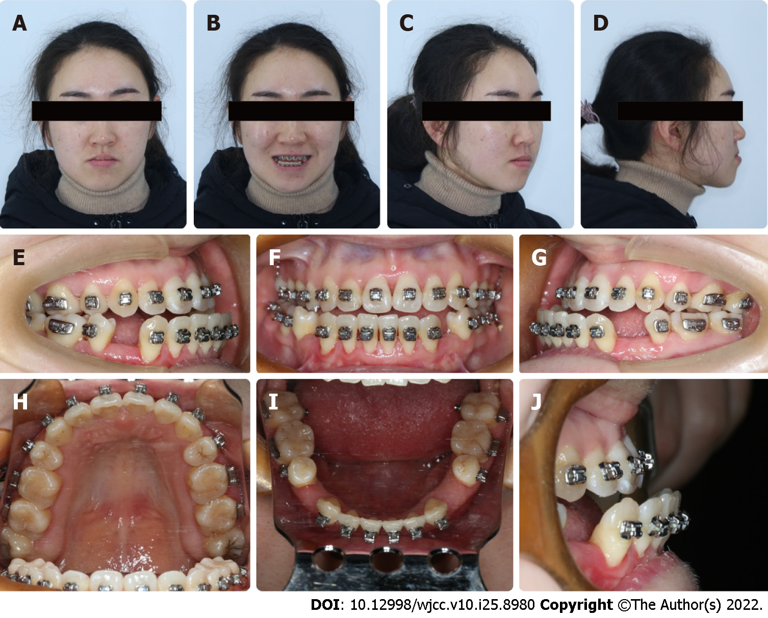
Figure 1 Extraoral and intraoral photographs.
A-D: Extraoral profiles showed facial asymmetry; E-J: Intraoral photographs showed dental malocclusion, thin periodontal phenotype and GR. GR: gingival recession.
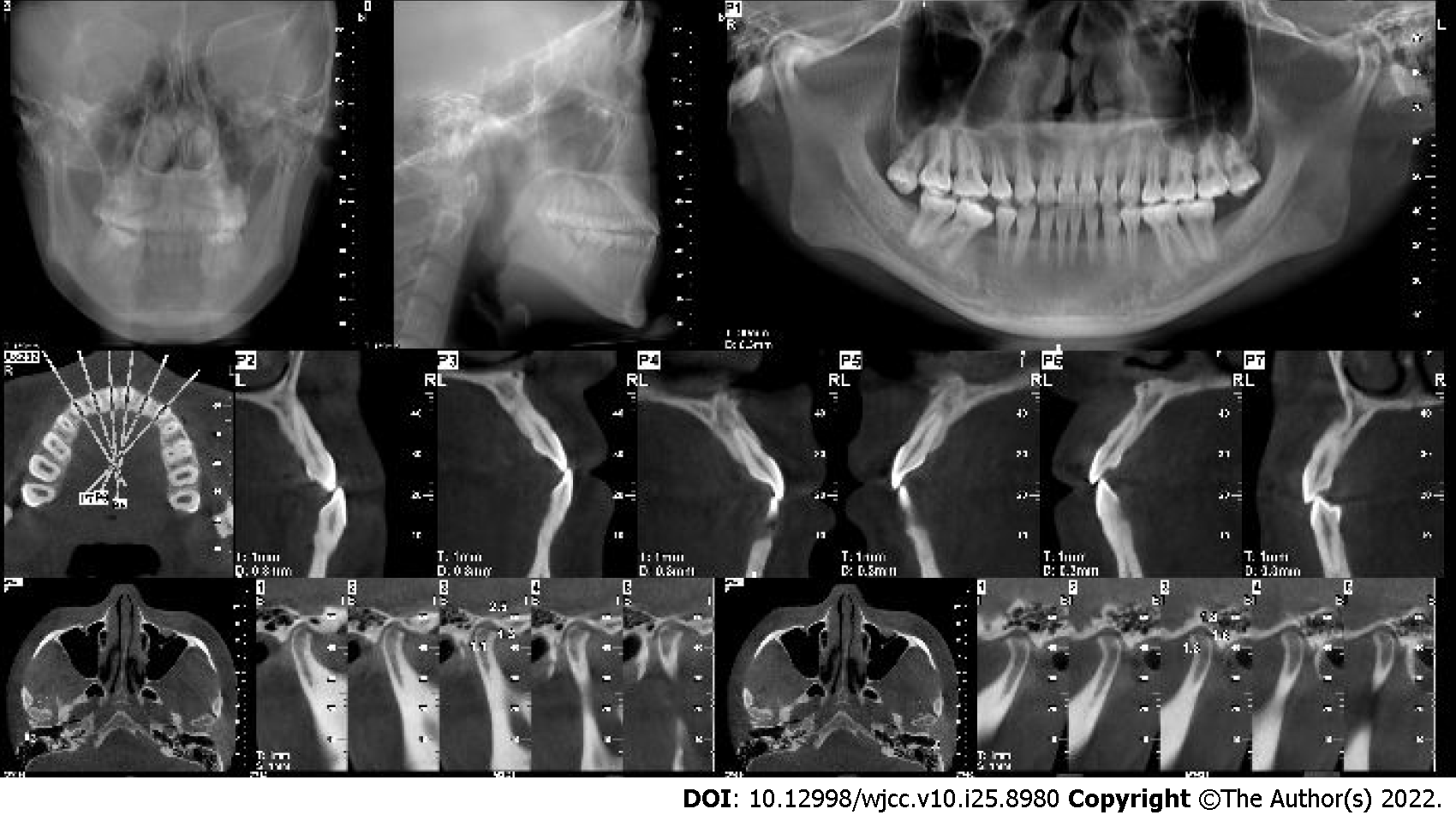
Figure 2 Cone-beam computed tomography.
Cone-beam computed tomography suggested thin alveolar bone morphology.
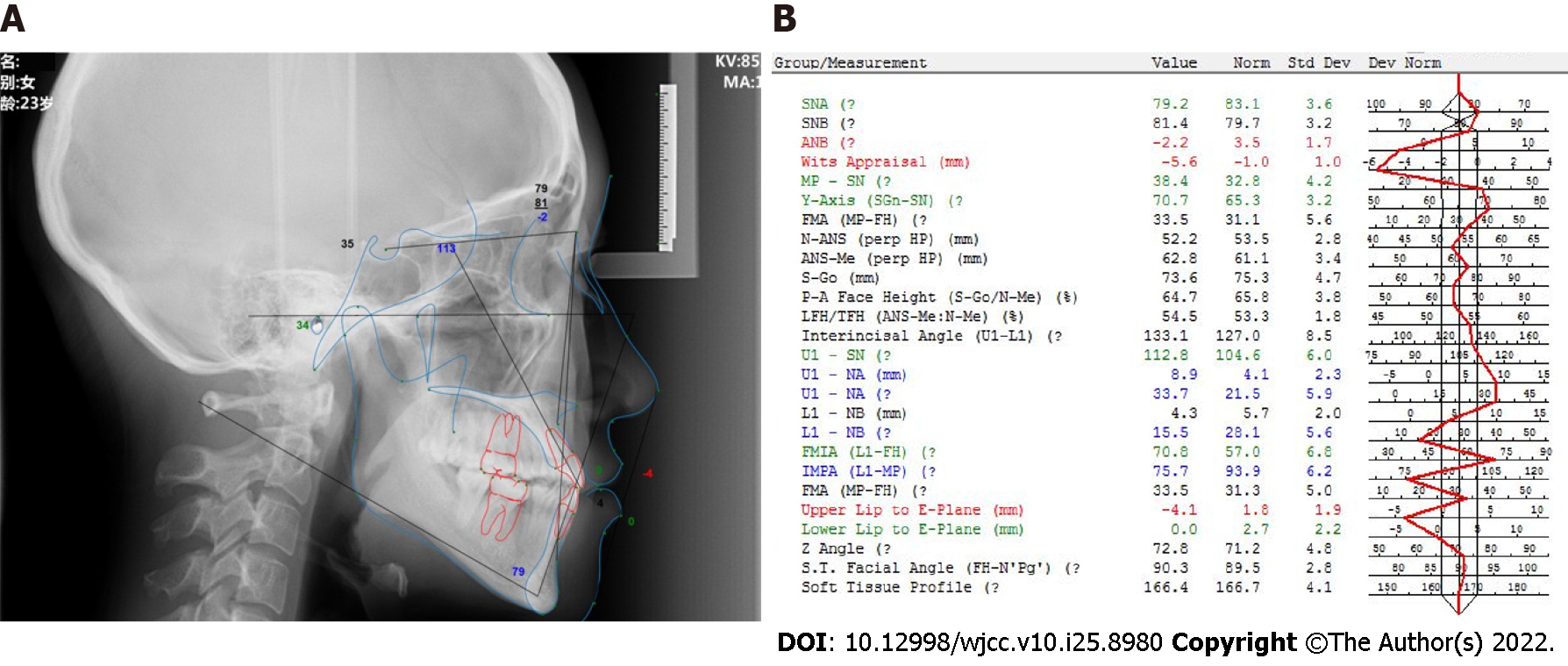
Figure 3 Cephalometric summary.
A: Cephalometric image; B: Cephalometric measurements.
Figure 4 Gingiva augmentation.
Mucograft was grafted to #33-#43. A: Before the operation; B: Suture; C: Three weeks after the procedure.
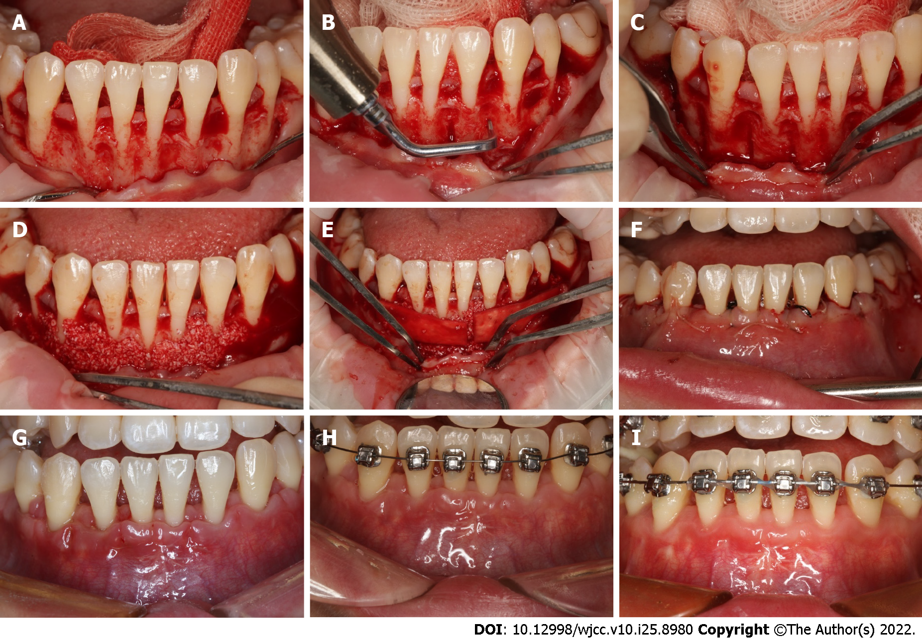
Figure 5 Surgical procedure for periodontal accelerated osteogenic orthodontics and postoperative follow-up.
A: Bone dehiscence can be observed after elevation of the full-thickness flap; B: The piezo surgery knife was used to perform decortication; C: Vertical alveolar decortication; D: The bone grafts were placed; E: The grafts were covered with an absorbable collagen membrane; F: Suture, the wound was well-closed; G: Image obtained two weeks after periodontal accelerated osteogenic orthodontics (PAOO); H: Three weeks after PAOO; I: Three months after PAOO; the postoperative images showed the wound healed well.
Figure 6 Completion of decompensation at 10 mo after periodontal accelerated osteogenic orthodontics.
A-D: Extraoral profiles; E-J: Intraoral photographs.
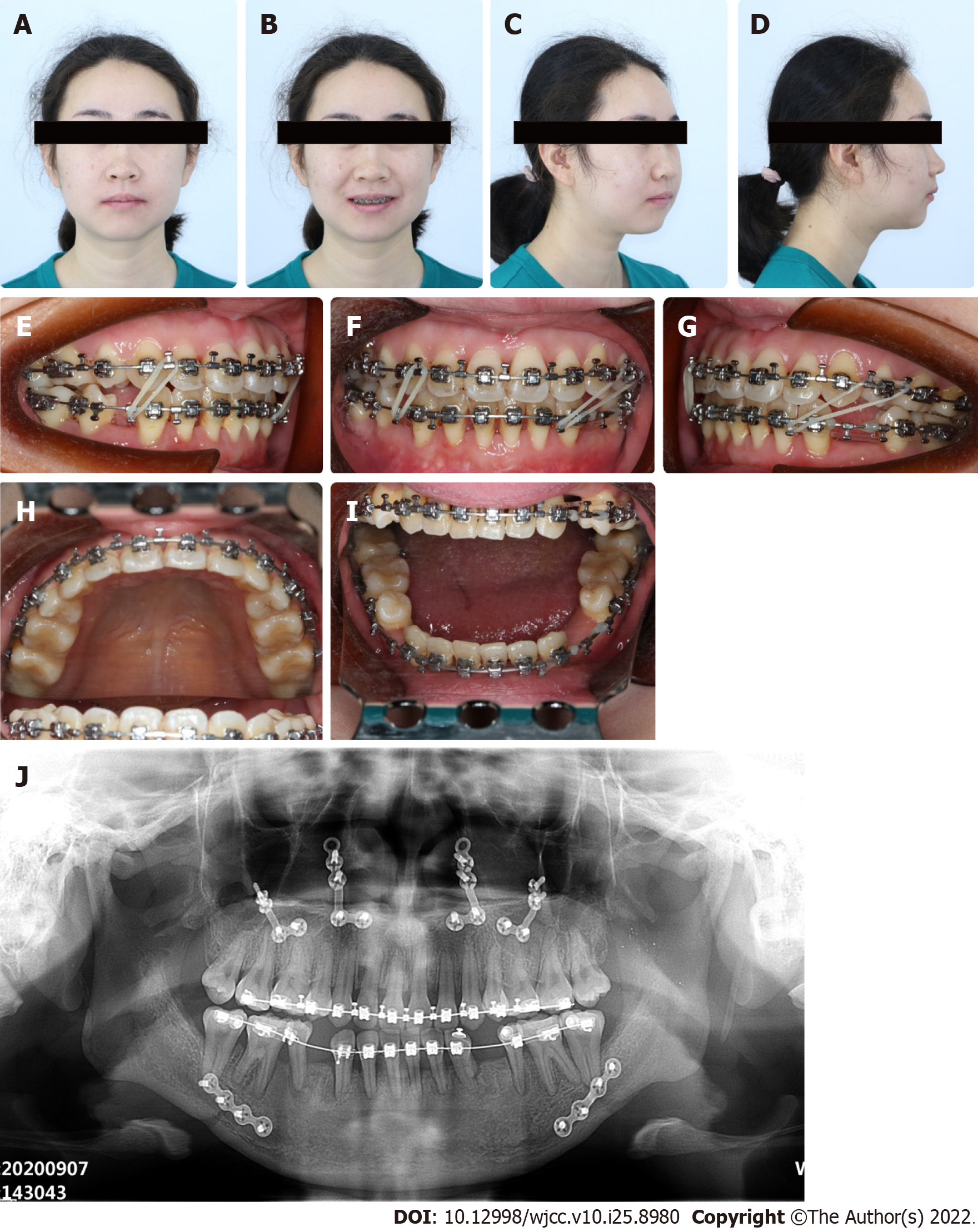
Figure 7 Five months after orthognathic surgery.
A-D: Extraoral profiles showing improvement of facial appearance; E-I: Intraoral photographs showing the correction of occlusion; J: Panoramic radiograph.
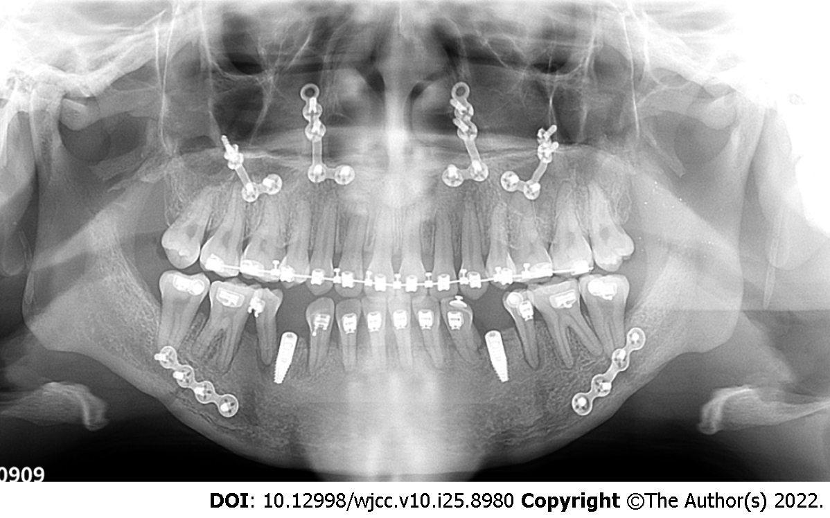
Figure 8
Panoramic radiograph after implant surgery.
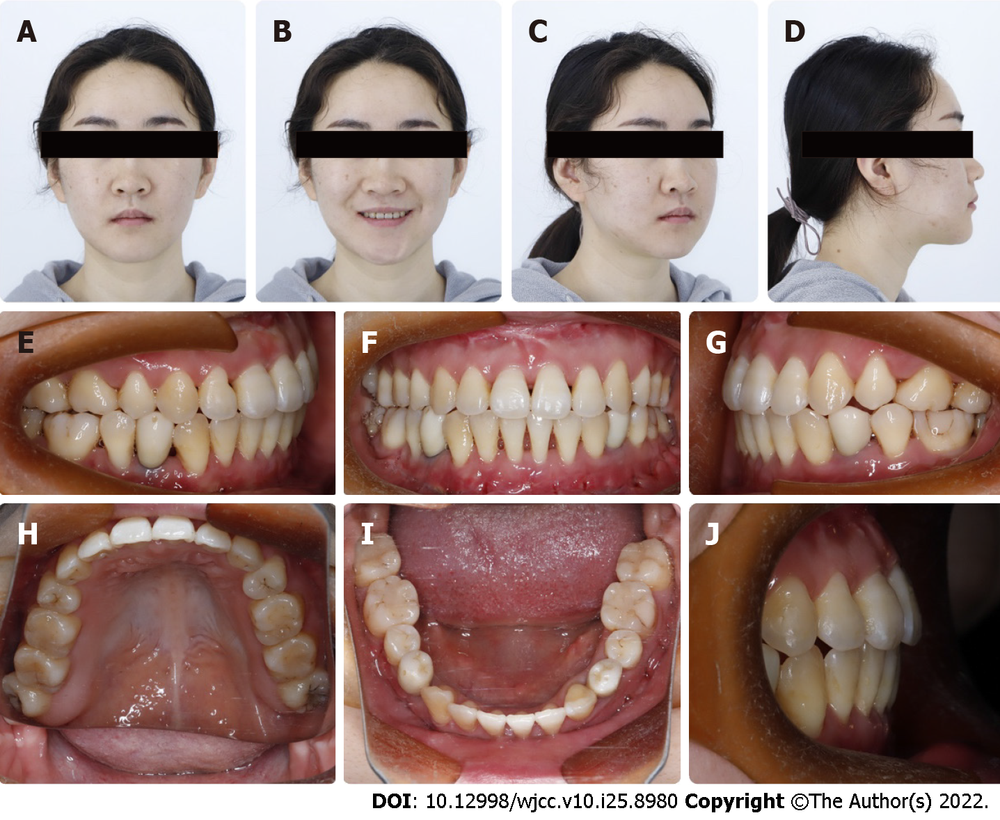
Figure 9 Completion of treatment.
A-D: Extraoral profiles; E-J: Intraoral photographs.
- Citation: Liu JY, Li GF, Tang Y, Yan FH, Tan BC. Multi-disciplinary treatment of maxillofacial skeletal deformities by orthognathic surgery combined with periodontal phenotype modification: A case report. World J Clin Cases 2022; 10(25): 8980-8989
- URL: https://www.wjgnet.com/2307-8960/full/v10/i25/8980.htm
- DOI: https://dx.doi.org/10.12998/wjcc.v10.i25.8980


