Copyright
©The Author(s) 2022.
World J Clin Cases. Aug 16, 2022; 10(23): 8417-8421
Published online Aug 16, 2022. doi: 10.12998/wjcc.v10.i23.8417
Published online Aug 16, 2022. doi: 10.12998/wjcc.v10.i23.8417
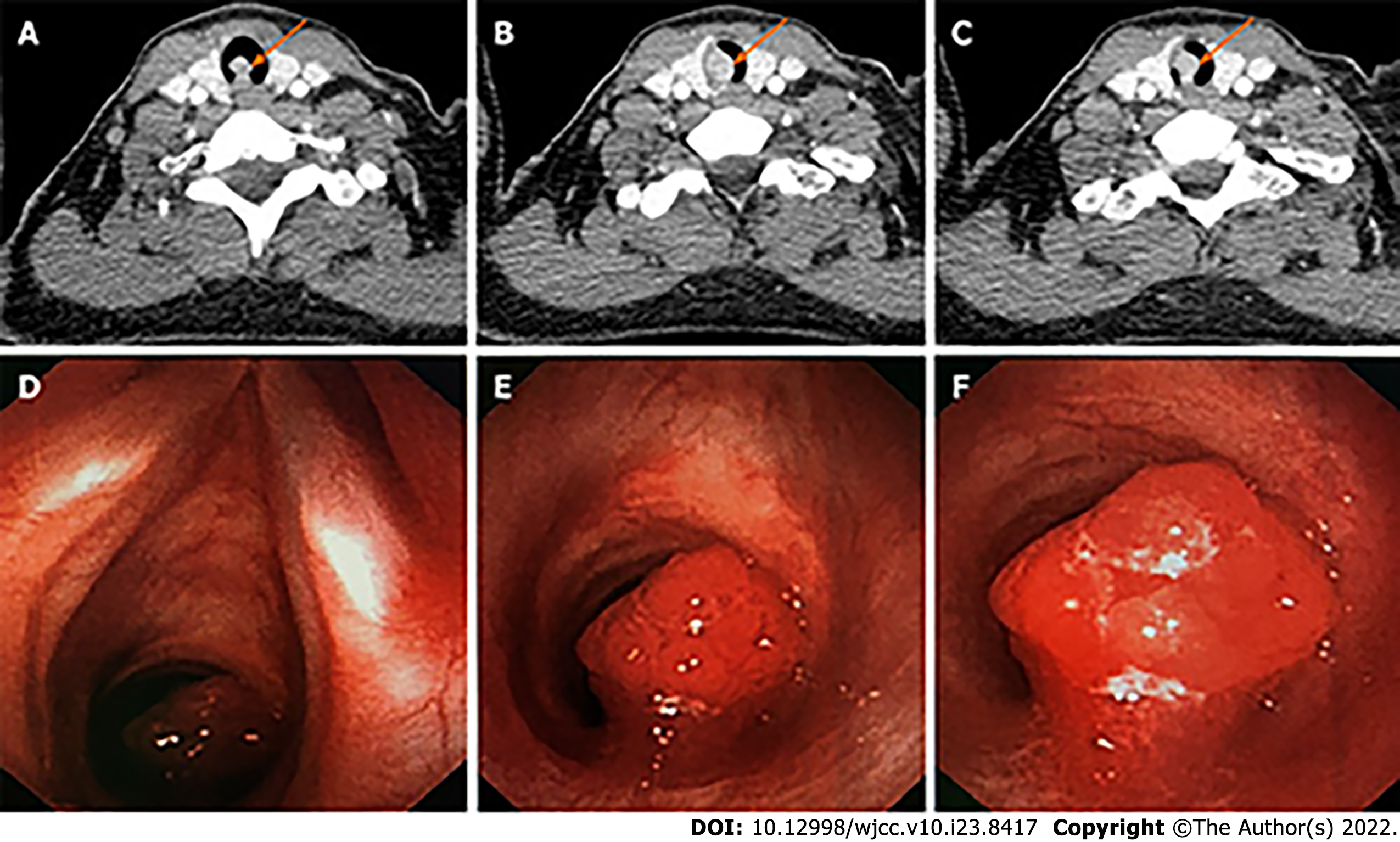
Figure 1 Neck computed tomography and bronchoscopic view.
A-C: Neck computed tomography showed a mass with soft-tissue density in the trachea pointed by orange arrows; D-F: In the bronchoscopic view, there was a mucosal neoplasm one cartilage ring below the glottis that occluded more than 90% of the lumen.
- Citation: Xu XH, Gao H, Chen XM, Ma HB, Huang YG. Using ketamine in a patient with a near-occlusion tracheal tumor undergoing tracheal resection and reconstruction: A case report. World J Clin Cases 2022; 10(23): 8417-8421
- URL: https://www.wjgnet.com/2307-8960/full/v10/i23/8417.htm
- DOI: https://dx.doi.org/10.12998/wjcc.v10.i23.8417


