Copyright
©The Author(s) 2022.
World J Clin Cases. Aug 16, 2022; 10(23): 8367-8374
Published online Aug 16, 2022. doi: 10.12998/wjcc.v10.i23.8367
Published online Aug 16, 2022. doi: 10.12998/wjcc.v10.i23.8367
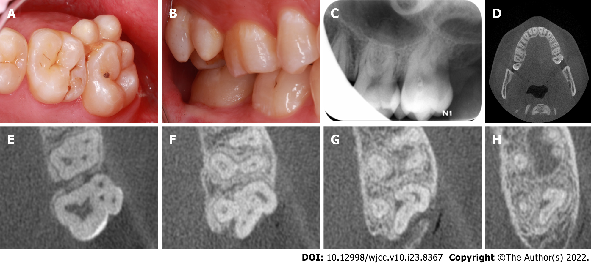
Figure 1 Representative preoperative intraoral photographs and radiographs of the fusion tooth.
A: Occlusal intraoral view revealing the presence of two supernumerary paramolars on the buccal aspect of the maxillary left second molar, with evidence of distinct developmental grooves separating these teeth and additional evidence of food impaction; B: Buccal view; C: A preoperative radiograph of the left maxillary second molar; D: Cone-beam computed tomography (CBCT) axial cross-sections of the maxillary fused second molar and paramolars; E-H: CBCT images revealed three slightly curved, patent root canals, which were associated with the maxillary second molar, and there was single similar canal for each of the fused paramolars. The apex of the irregular bending isthmus area that was formed by the intersection of three teeth was connected in a periapical transmission image.
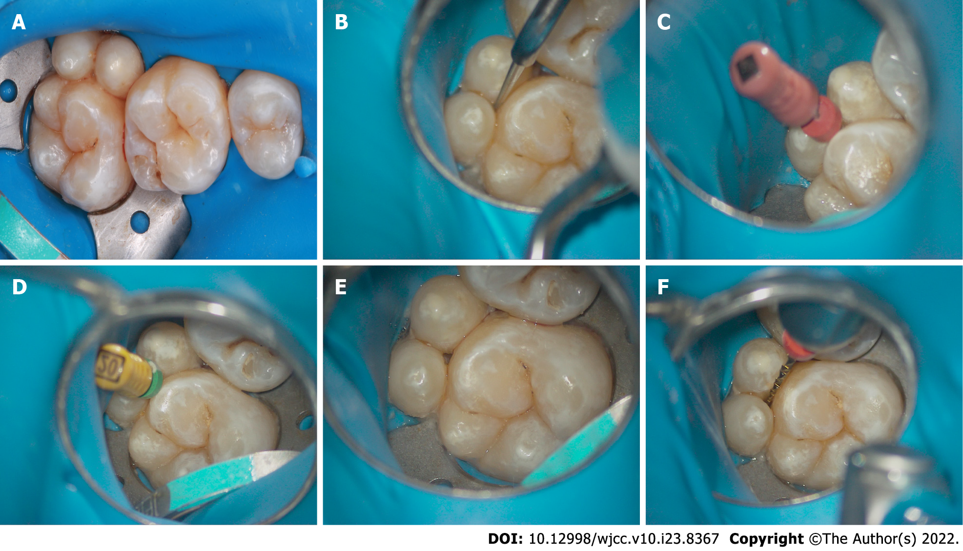
Figure 2 Representative intraoral photos of the fused tooth during root canal therapy.
A: A rubber dam was positioned; B: A ball drill (ET BD) and ET 20 of the ultrasonic equipment (P5 Newtron) were used to remove the damaged tissue under a microscope; C: An electronic apex locator was utilized to determine the working lengths with 6#K-file; D-E: Filing was performed until size 20# was reached; F: Waveone Gold Ni-Ti rotary instruments were used to clean and shape the canals.
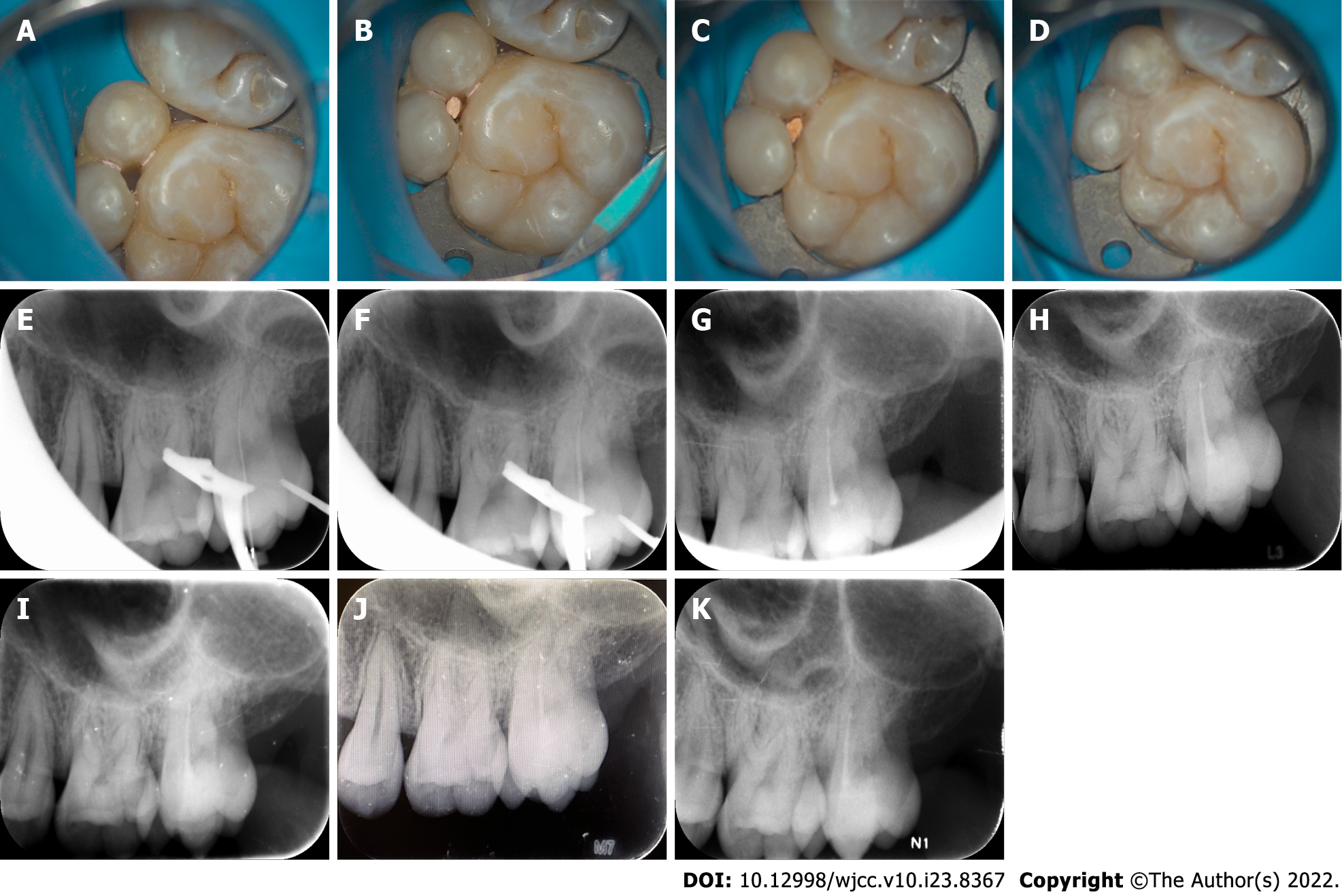
Figure 3 Representative intraoral photos of the fused tooth during root canal therapy and radiographs at different follow-up periods.
A: The canals were dried following a final irrigation; B: Occlusal view of the canal tip; C: Complete canal obturation was achieved via vertical gutta-percha condensation; D: Image of the nanoresin-mediated seal of the access cavity; E: Working length radiograph; F: Cone fit radiograph; G: A final digital PAX revealed that the canals were well-obturated; H-K: A radiograph taken during the 6-, 12-, 18-, 24-mo follow-ups revealed good treatment outcomes.
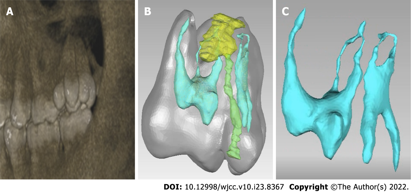
Figure 4 The construction of digital model of the fused tooth's local anatomy.
A-C: A digital model of the local anatomy was constructed, revealing that the roots of the two supernumerary teeth were fused with the mesial buccal roots of the second molar, and the root canal systems of the two supernumerary teeth intersected the middle 1/3 of the root and apical area while remaining separated from the second molar root canal system.
- Citation: Mei XH, Liu J, Wang W, Zhang QX, Hong T, Bai SZ, Cheng XG, Tian Y, Jiang WK. Endodontic management of a fused left maxillary second molar and two paramolars using cone beam computed tomography: A case report. World J Clin Cases 2022; 10(23): 8367-8374
- URL: https://www.wjgnet.com/2307-8960/full/v10/i23/8367.htm
- DOI: https://dx.doi.org/10.12998/wjcc.v10.i23.8367


