Copyright
©The Author(s) 2022.
World J Clin Cases. Aug 16, 2022; 10(23): 8323-8329
Published online Aug 16, 2022. doi: 10.12998/wjcc.v10.i23.8323
Published online Aug 16, 2022. doi: 10.12998/wjcc.v10.i23.8323
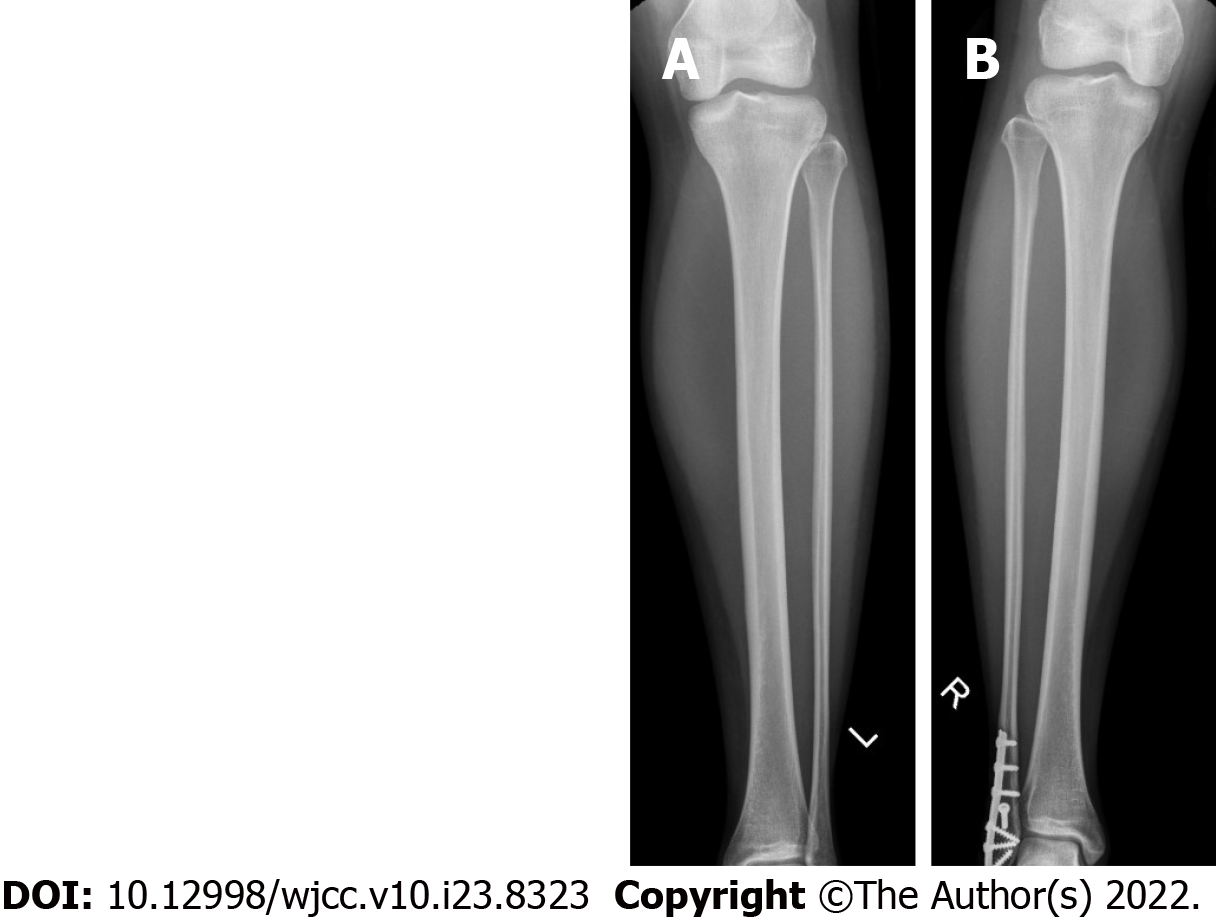
Figure 1 Radiograph.
A: Radiograph of left tibia; B: Radiograph of right tibia (previous healed fibula fracture fixation).
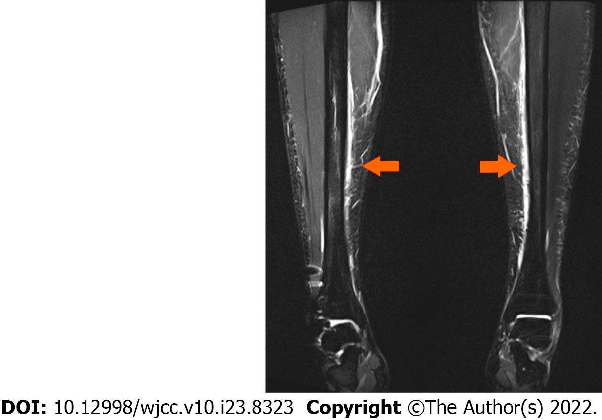
Figure 2 Magnetic resonance imaging bilateral tibiae.
Arrows denote regions of periosteal oedema.
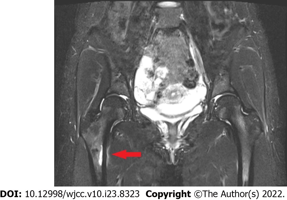
Figure 3 Magnetic resonance imaging pelvis.
Arrow demonstrates stress fracture right medial subtrochanteric region.
- Citation: Tan DS, Cheung FM, Ng D, Cheung TLA. Femoral neck stress fracture and medial tibial stress syndrome following high intensity interval training: A case report and review of literature. World J Clin Cases 2022; 10(23): 8323-8329
- URL: https://www.wjgnet.com/2307-8960/full/v10/i23/8323.htm
- DOI: https://dx.doi.org/10.12998/wjcc.v10.i23.8323


