Copyright
©The Author(s) 2022.
World J Clin Cases. Aug 16, 2022; 10(23): 8312-8322
Published online Aug 16, 2022. doi: 10.12998/wjcc.v10.i23.8312
Published online Aug 16, 2022. doi: 10.12998/wjcc.v10.i23.8312
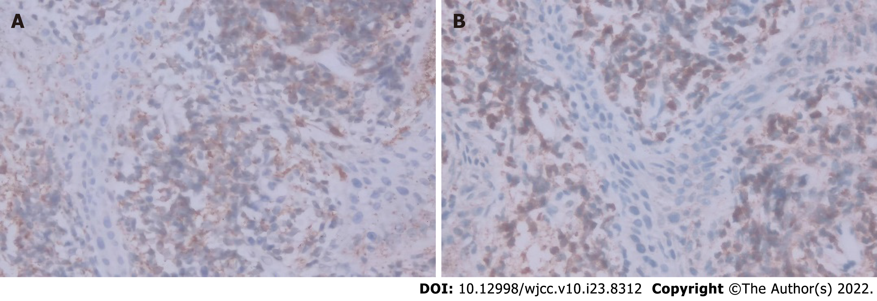
Figure 1 Immunohistochemistry of vaginal biopsy.
A: CD99 positive staining (× 400); B: Myeloperoxidase positive staining (× 400) were exhibited in tumor cells.
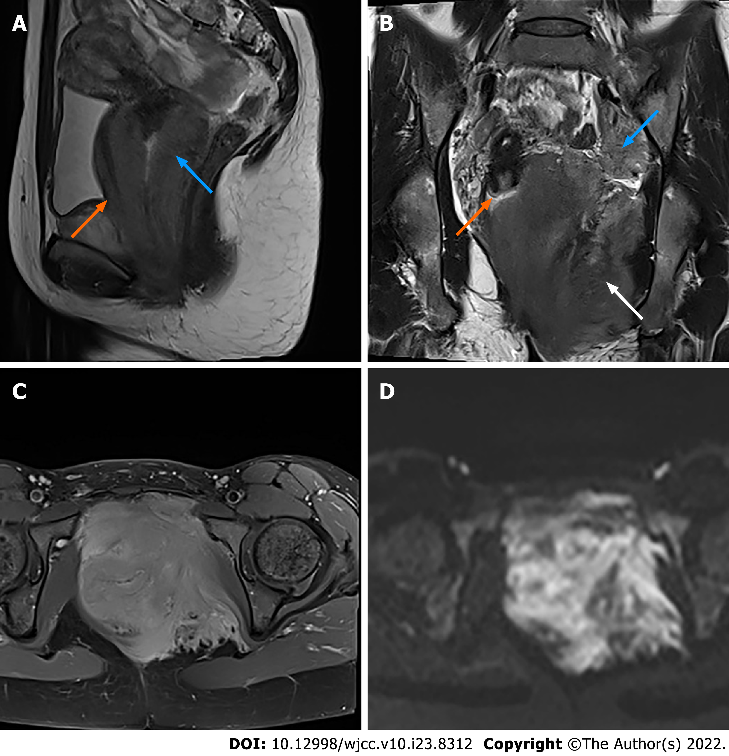
Figure 2 Magnetic resonance imaging with contrast enhancement of the pelvis.
A: Sagittal T2WI shows a diffuse mass of the vaginal wall (blue arrow) with involvement of the posterior wall of the urethra (orange arrow); B: Coronal T2WI shows massive infiltration of the left pelvic floor (white arrow), while the cervix was intact (orange arrow). The left obturator lymph node was enlarged (blue arrow); C: Axial contrast-enhanced T1WI shows that the lesion was homogeneously enhanced, greater than the muscle; D: On diffusion-weighted imaging (b = 800), the lesion was hyperintense.
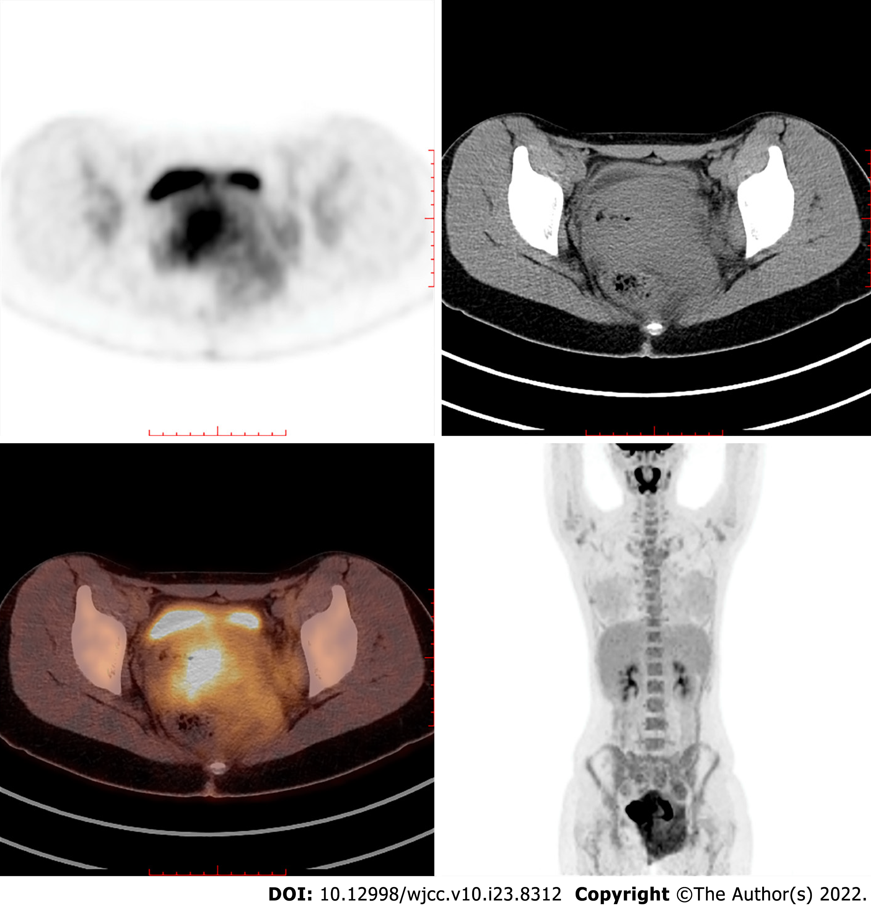
Figure 3 Positron emission tomography-computed tomography shows high fluorodeoxyglucose uptake of the mass and the enlarged left obturator lymph node.
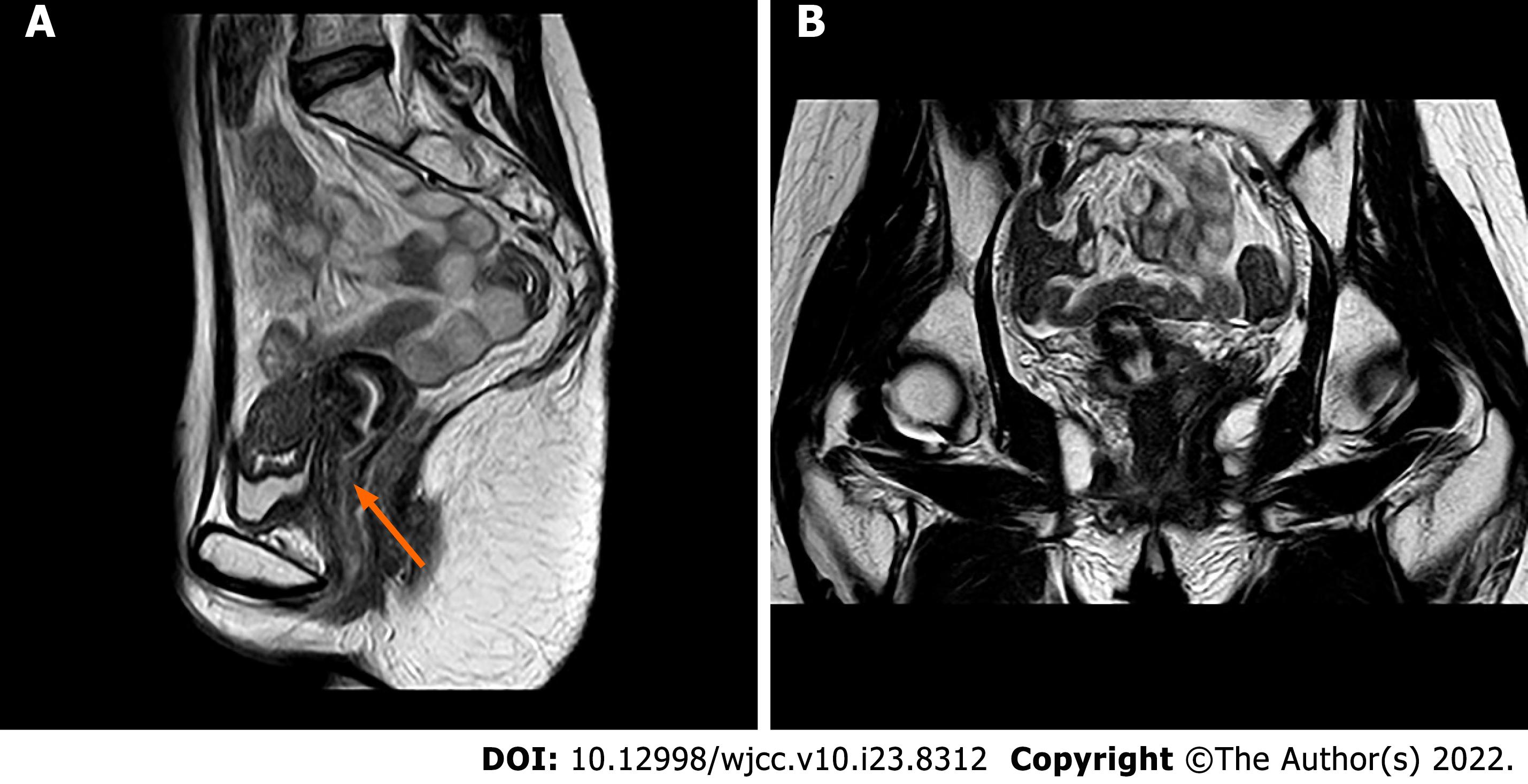
Figure 4 One year after complete remission, pelvic magnetic resonance imaging shows no sign of tumour recurrence on either the vagina (orange arrow in A) or the pelvic floor (B).
- Citation: Wang JX, Zhang H, Ning G, Bao L. Vulvovaginal myeloid sarcoma with massive pelvic floor infiltration: A case report and review of literature. World J Clin Cases 2022; 10(23): 8312-8322
- URL: https://www.wjgnet.com/2307-8960/full/v10/i23/8312.htm
- DOI: https://dx.doi.org/10.12998/wjcc.v10.i23.8312


