Copyright
©The Author(s) 2022.
World J Clin Cases. Aug 16, 2022; 10(23): 8262-8270
Published online Aug 16, 2022. doi: 10.12998/wjcc.v10.i23.8262
Published online Aug 16, 2022. doi: 10.12998/wjcc.v10.i23.8262
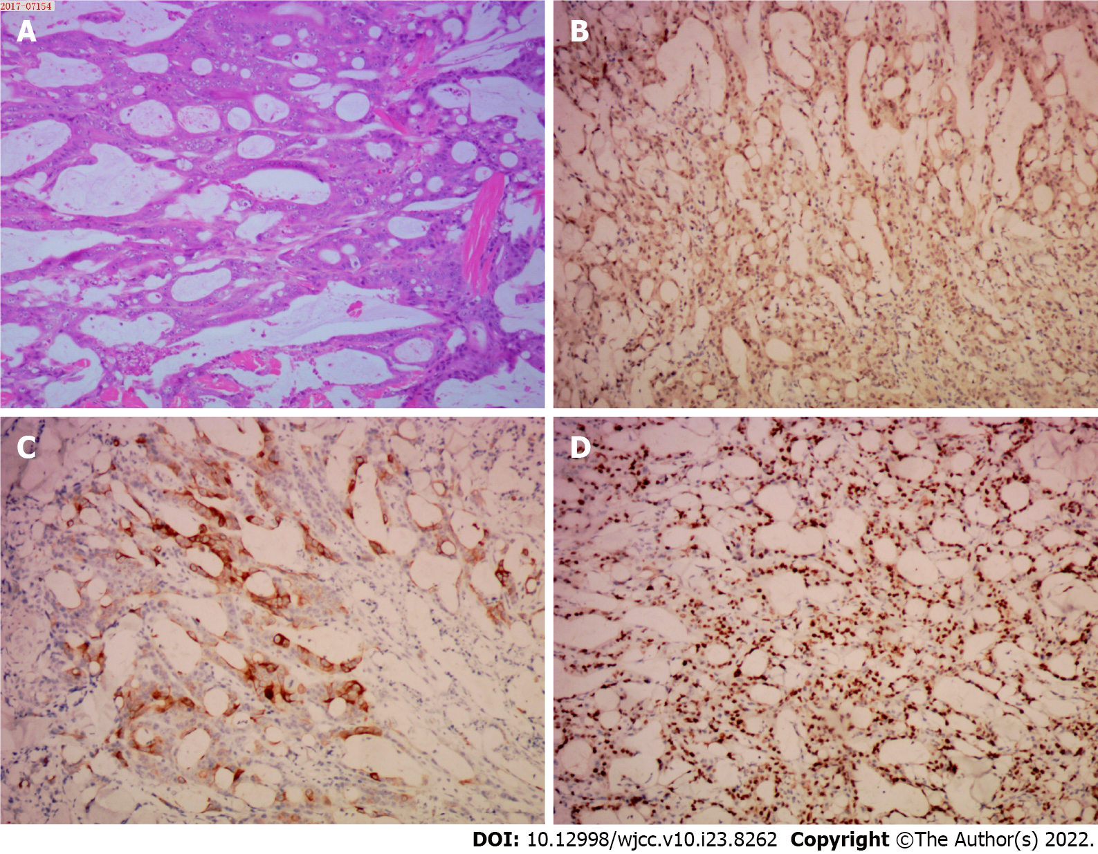
Figure 1 The pathological examination of the left shoulder cutaneous mass.
The hematoxylin and eosin and immunohistochemical staining of resected specimen at × 10 magnification. A: Hematoxylin and eosin; B: CDX2; C: CK20; D: Ki67.
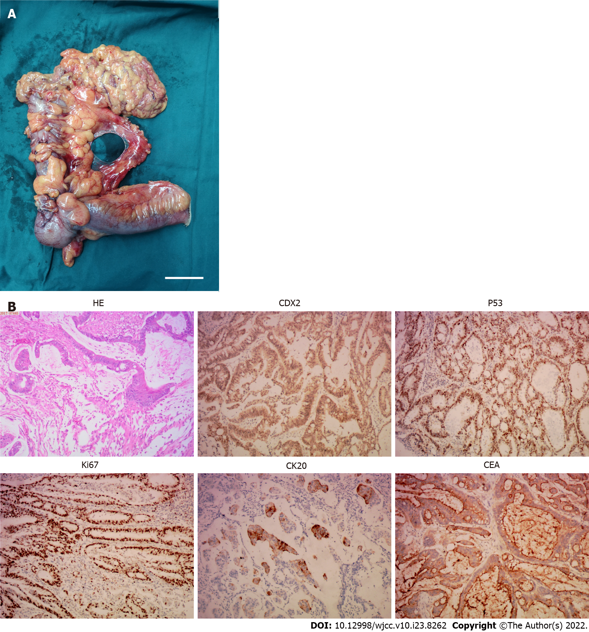
Figure 2 The pathological examination of the ascending colon tumor mass.
A: The resected tumor mass, the scale bar represents 5 cm; B: The hematoxylin and eosin and immunohistochemical staining of resected specimen at × 10 magnification. HE: Hematoxylin and eosin; CEA: Carcinoembryonic antigen.
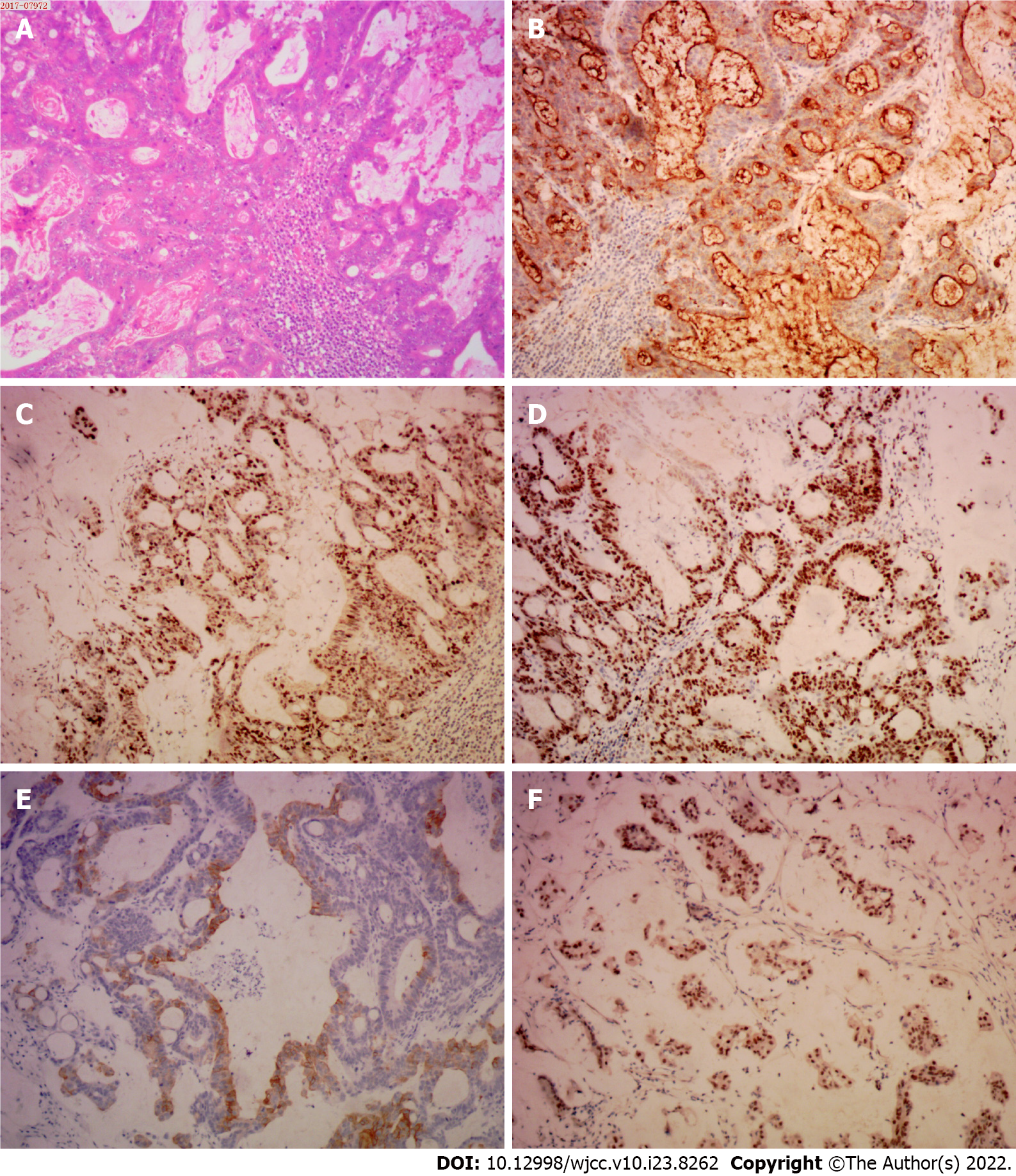
Figure 3 The pathological examination of the cervical lymph node.
The hematoxylin and eosin and immunohistochemical staining of resected specimen at × 10 magnification. A: Hematoxylin and eosin; B: Carcinoembryonic antigen; C: P53; D: Ki67; E: CK20; F: CDX2.
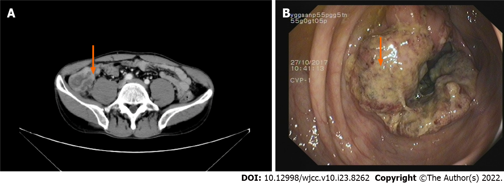
Figure 4 The image examination of the abdomen and colonoscopy.
A: Enhanced computed tomography examination of the abdomen; the orange arrow indicates thickening and edema of the ascending colon (ileocecal region); B: Colonoscopy revealed a cauliflower-like mass in the ascending colon; the orange arrow indicates the ascending colon tumor mass.
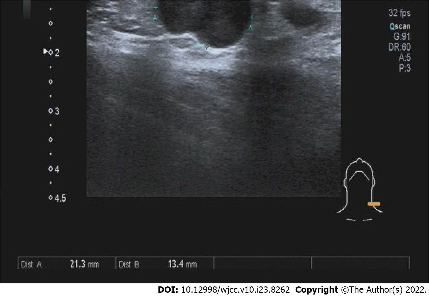
Figure 5 The color Doppler ultrasound image of the left neck.
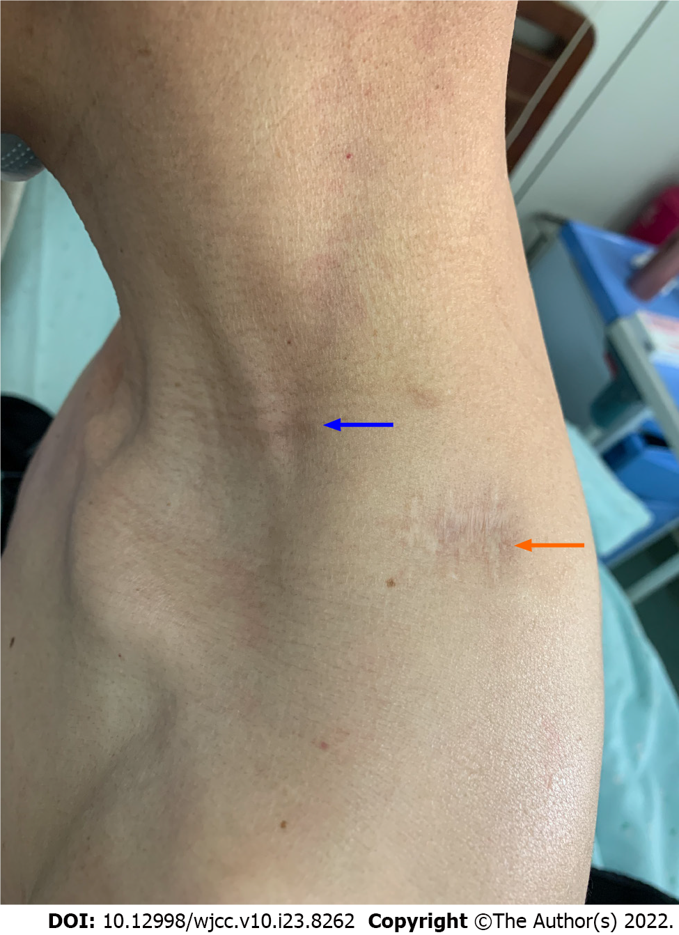
Figure 6 The image of recovered surgical wounds.
The orange arrow indicated the wound of resected left shoulder cutaneous mass. The blue arrow indicated the wound of resected cervical lymph nodes.
- Citation: Zhou JC, Wang JJ, Liu T, Tong Q, Fang YJ, Wu ZQ, Hong Q. Primary ascending colon cancer accompanying skip metastases in left shoulder skin and left neck lymph node: A case report. World J Clin Cases 2022; 10(23): 8262-8270
- URL: https://www.wjgnet.com/2307-8960/full/v10/i23/8262.htm
- DOI: https://dx.doi.org/10.12998/wjcc.v10.i23.8262


