Copyright
©The Author(s) 2022.
World J Clin Cases. Aug 16, 2022; 10(23): 8255-8261
Published online Aug 16, 2022. doi: 10.12998/wjcc.v10.i23.8255
Published online Aug 16, 2022. doi: 10.12998/wjcc.v10.i23.8255
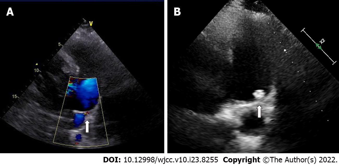
Figure 1 Patent ductus arteriosus assessment by transthoracic echocardiography.
A: A 5.1-mm patent ductus arteriosus before occlusion (arrow); B: No residual shunt 2 years after occlusion (arrow).
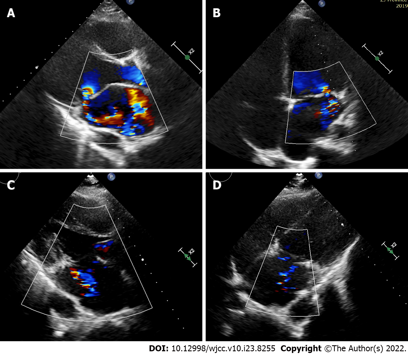
Figure 2 Mitral valve assessment by transthoracic echocardiography.
A and B: Echocardiography before surgery showed mitral anterior leaflet prolapse with severe eccentric regurgitation; C and D: Two years after surgery, echocardiography showed mild mitral regurgitation.
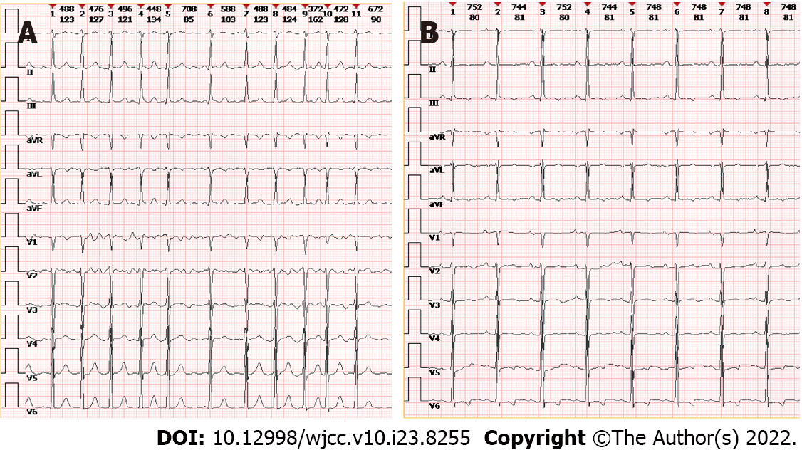
Figure 3 Electrocardiogram.
A: Baseline; B: Postsurgery.
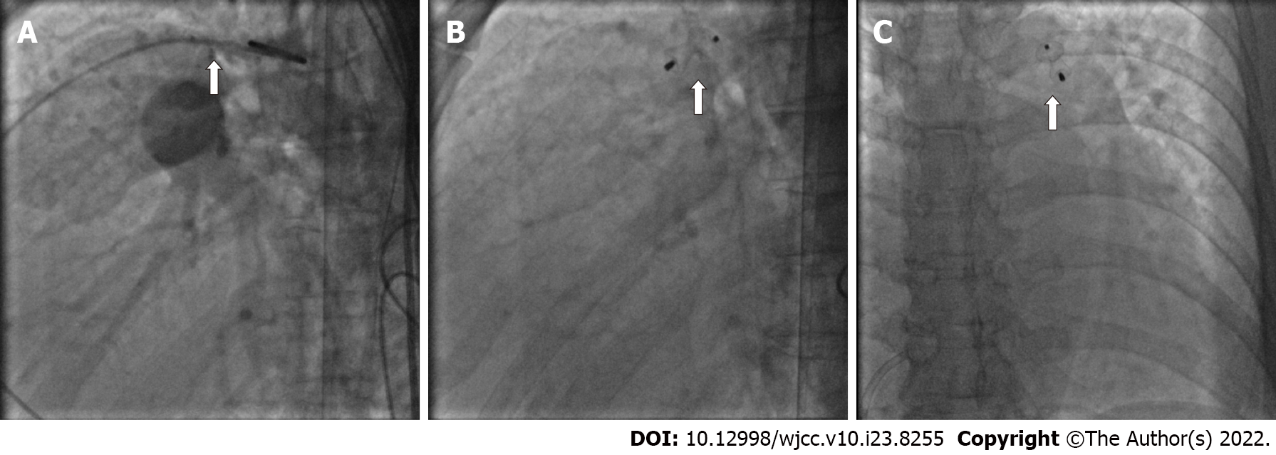
Figure 4 Transcatheter patent ductus arteriosus occlusion.
A: Patent ductus arteriosus (PDA) in the catheterization (arrow); B and C: complete closure of PDA during catheterization and the PDA occluder (arrow).
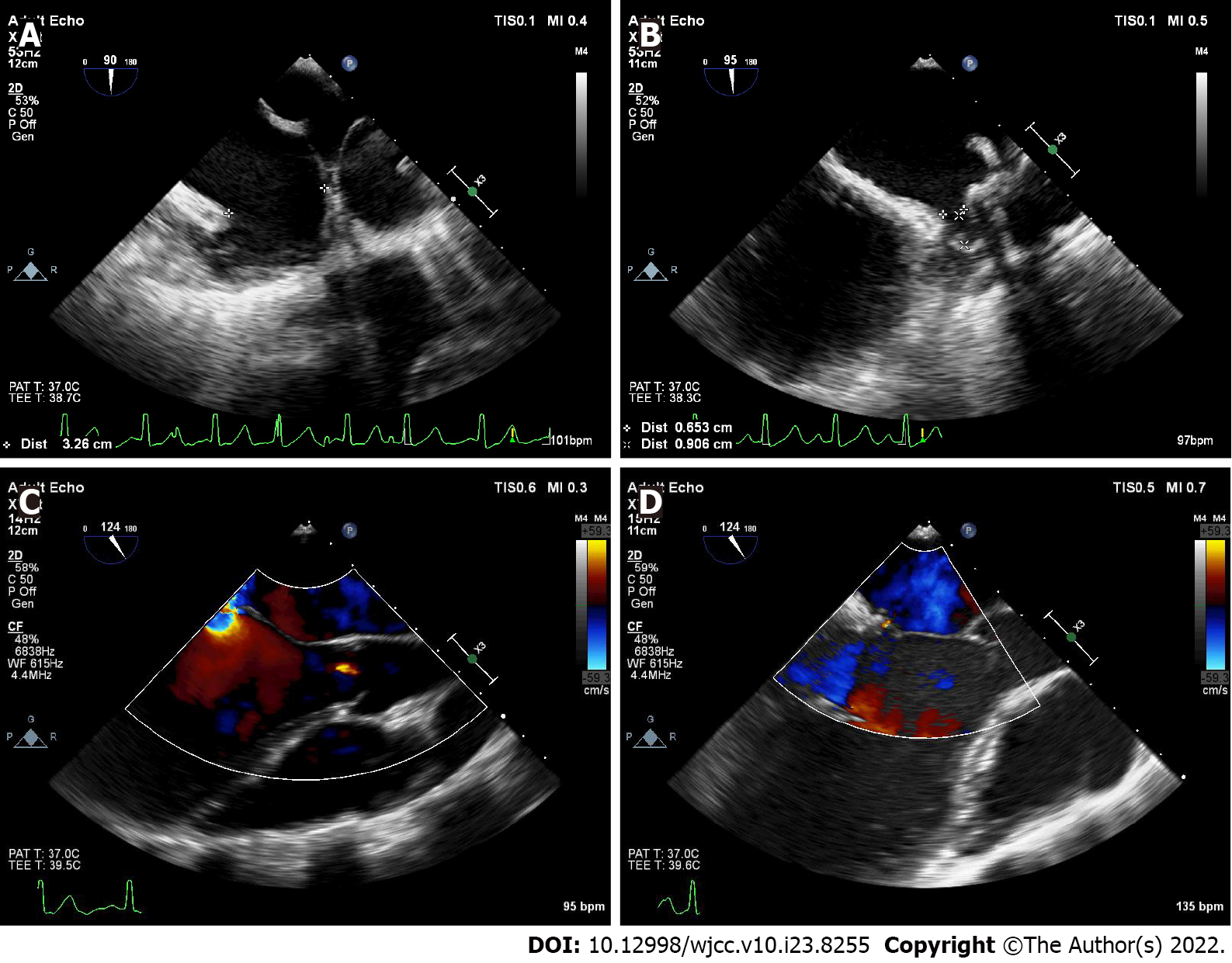
Figure 5 Intraoperative transesophageal echocardiography.
A: Left atrial appendage assessment; B: Left atrial appendage occlusion; C: Severe eccentric mitral regurgitation before chordal replacement; D: Decreased mitral regurgitation after chordal replacement.
- Citation: Zhao CZ, Yan Y, Cui Y, Zhu N, Ding XY. Sequential multidisciplinary minimally invasive therapeutic strategy for heart failure caused by four diseases: A case report. World J Clin Cases 2022; 10(23): 8255-8261
- URL: https://www.wjgnet.com/2307-8960/full/v10/i23/8255.htm
- DOI: https://dx.doi.org/10.12998/wjcc.v10.i23.8255


