Copyright
©The Author(s) 2022.
World J Clin Cases. Aug 16, 2022; 10(23): 8232-8241
Published online Aug 16, 2022. doi: 10.12998/wjcc.v10.i23.8232
Published online Aug 16, 2022. doi: 10.12998/wjcc.v10.i23.8232
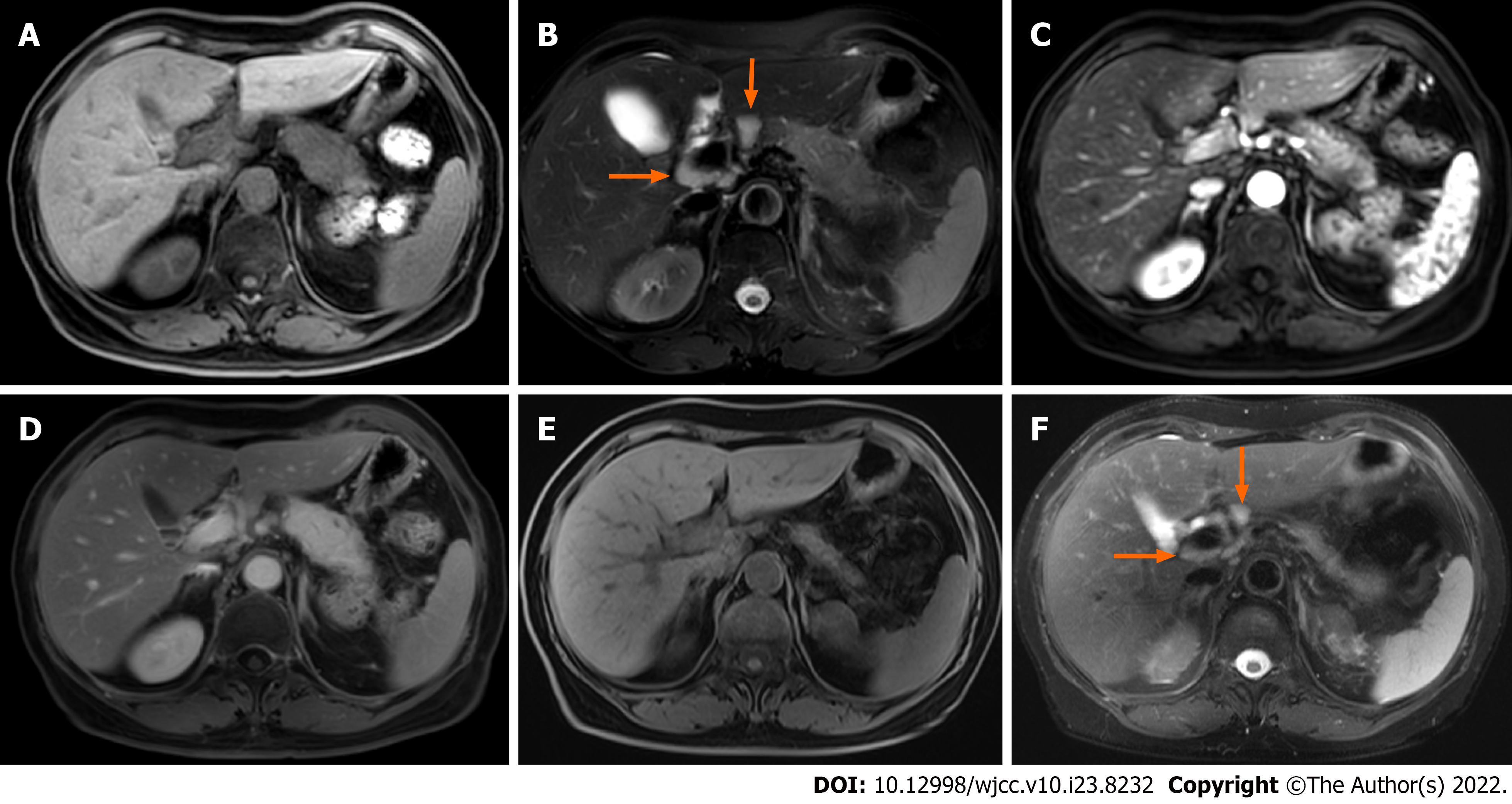
Figure 1 Focal autoimmune pancreatitis with atrophy after spontaneous remission in case 1.
A and B: The pancreatic body and tail enlarge, and multiple enlarged lymph nodes (arrows) are seen around the portal vein; C and D: Gradual enhancement of the lesion on enhanced scan; E and F: Magnetic resonance imaging follow-up at the 13th month. Pancreatic swelling dissipates and the volume of the pancreas decreases significantly, and the enlarged lymph nodes (arrows) are significantly reduced as well.
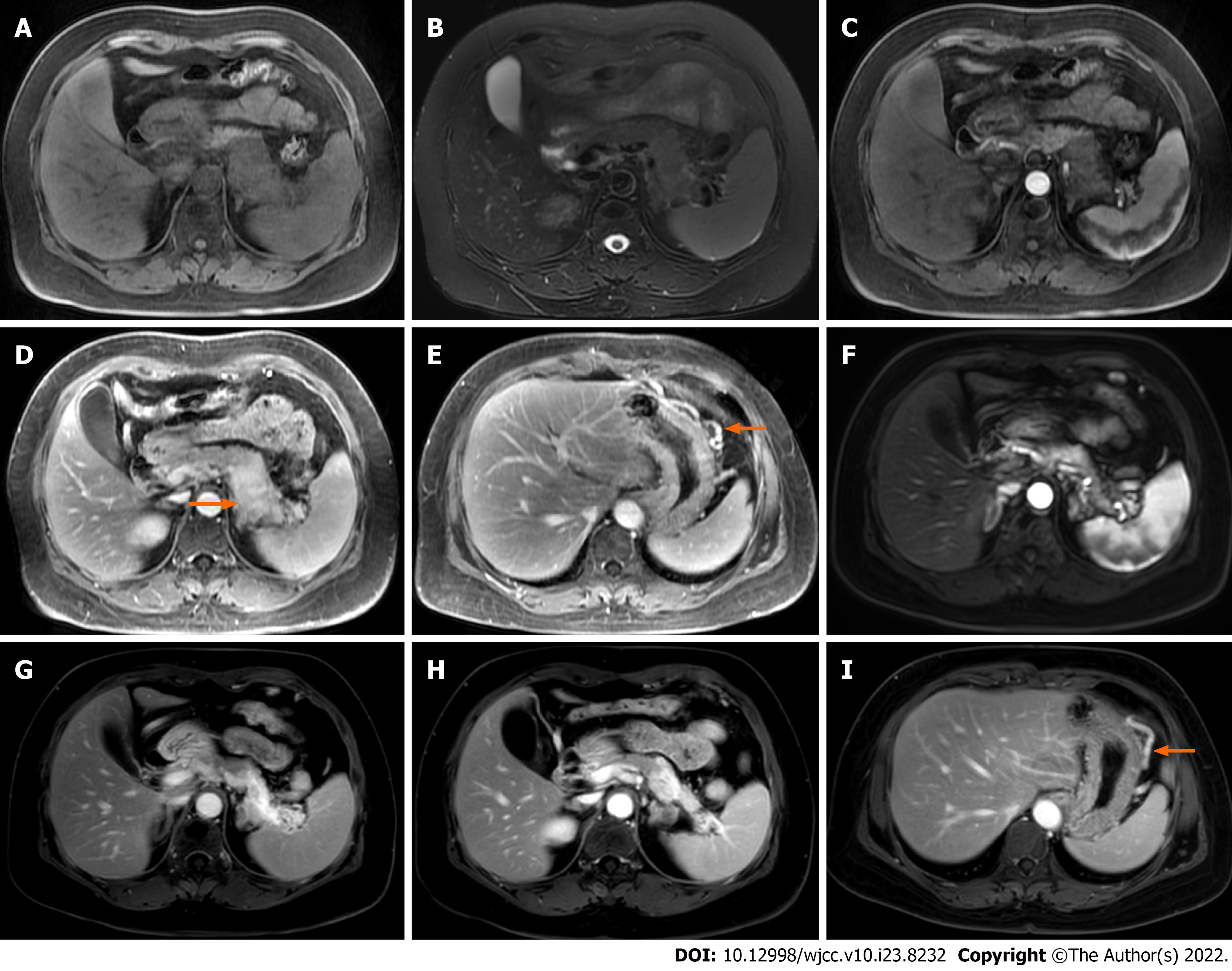
Figure 2 Focal autoimmune pancreatitis with progressive fibrosis after spontaneous remission in case 2.
A and B: Enlargement of the pancreatic body and tail; C, D, and E: In addition to the gradual enhancement of the lesion in the body and tail of the pancreas, stenosis of the splenic vein (arrow) and dilation of the large curved side vein of the gastric body (arrow) are observed; F and G: Magnetic resonance imaging (MRI) follow-up at 22 mo showing that the body and tail of the pancreas is significantly reduced and more obviously gradually enhanced; H: Lower level MRI showing that the volume of the pancreatic body and tail is significantly atrophied and obviously enhanced; I: The dilation of the large curvature side vein of the gastric body (arrow) is not improved.
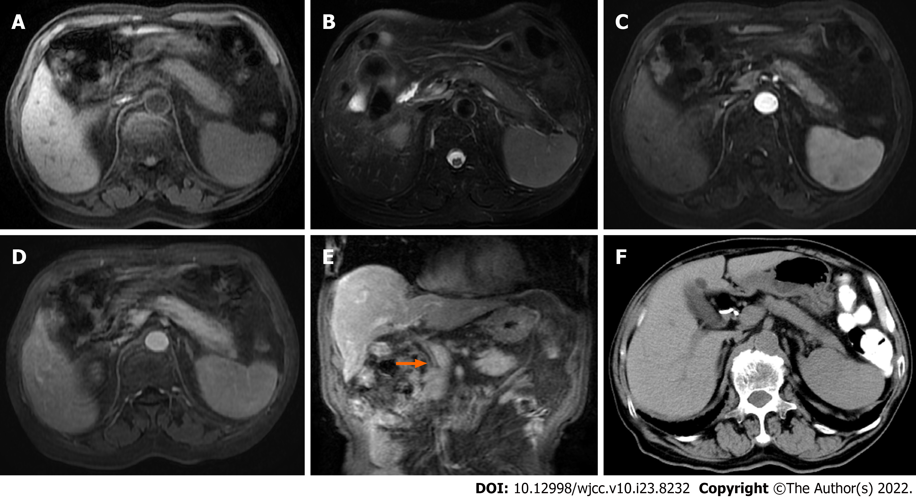
Figure 3 Diffuse autoimmune pancreatitis with spontaneous remission in case 3.
A and B: Diffuse enlargement of the pancreas; C and D: Progressive enhancement of the pancreatic lesion and the pseudocapsule on enhanced scan; E: Extra-hepatic bile duct wall thickening and enhancement (arrow); F: Computed tomography follow-up at 20 mo showing that the pancreatic swelling has disappeared.
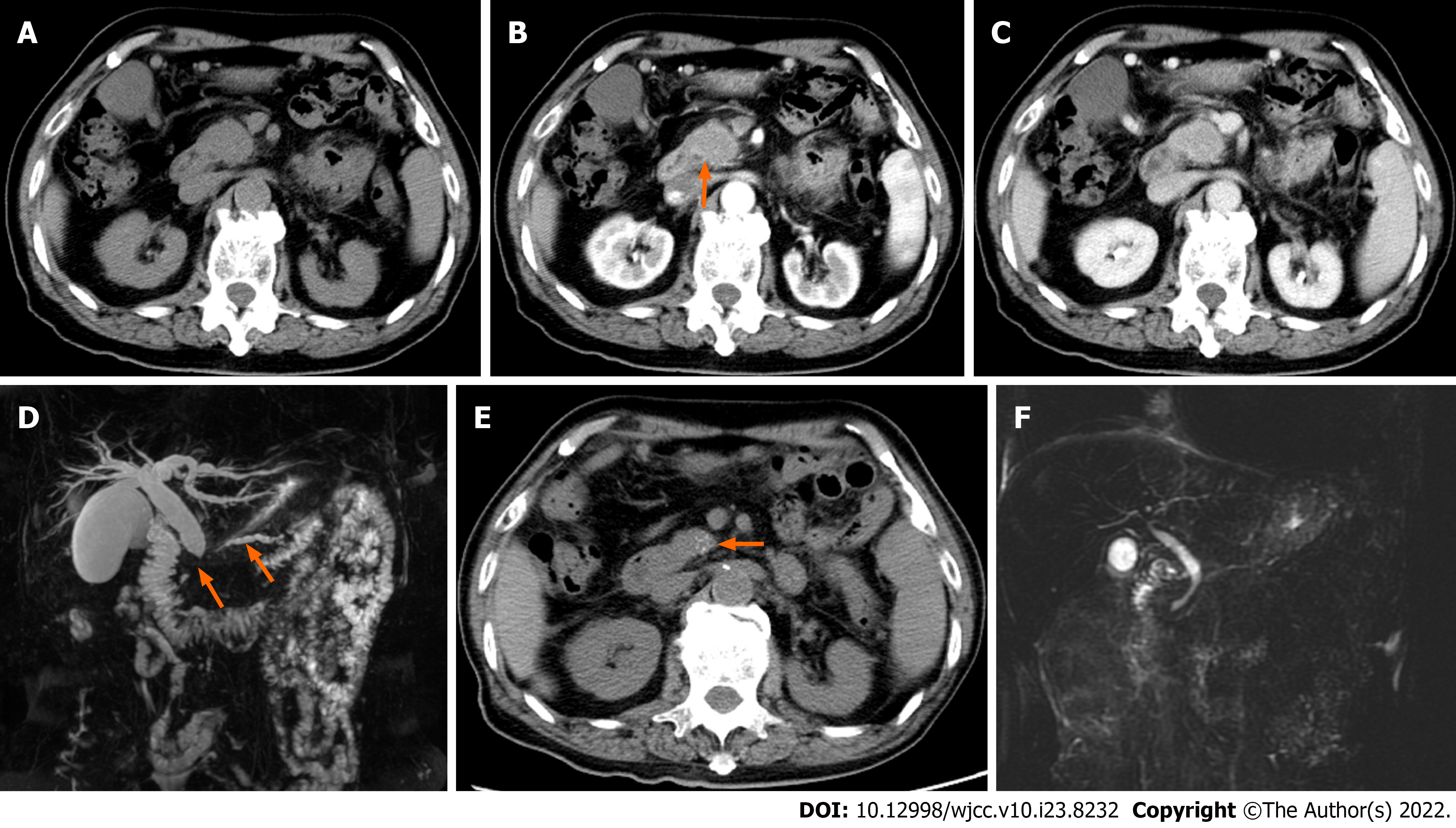
Figure 4 Focal autoimmune pancreatitis with calcification after spontaneous remission in case 4.
A, B, and C: The pancreatic head is enlarged with progressive enhancement, the wall of the common bile duct is thickened and abnormally enhanced (arrow); D: Magnetic resonance cholangiopancreatography (MRCP) showed a longer stenotic segment of the pancreatic duct (arrow) and bile duct, and dilatation of the upstream pancreatic duct (arrow) and proximal bile duct; E: Computed tomography follow-up after 12 mo showing that the swelling of the pancreatic head has dissipated, and multiple punctate calcifications (arrow) occur; F: The diseased pancreatic duct and bile ducts have nearly returned to normal on MRCP follow-up.
- Citation: Zhang BB, Huo JW, Yang ZH, Wang ZC, Jin EH. Spontaneous remission of autoimmune pancreatitis: Four case reports . World J Clin Cases 2022; 10(23): 8232-8241
- URL: https://www.wjgnet.com/2307-8960/full/v10/i23/8232.htm
- DOI: https://dx.doi.org/10.12998/wjcc.v10.i23.8232


