Copyright
©The Author(s) 2022.
World J Clin Cases. Aug 16, 2022; 10(23): 8115-8123
Published online Aug 16, 2022. doi: 10.12998/wjcc.v10.i23.8115
Published online Aug 16, 2022. doi: 10.12998/wjcc.v10.i23.8115
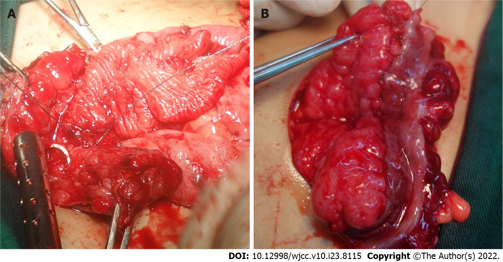
Figure 1 Intraoperative photos.
A: The whole layer intestinal patch was sutured with the bladder edge in the standard cystoplasty procedure; B: The double-layer intestine seromuscular patch was sutured with the bladder incisal edge in the seromuscular bladder augmentation procedure.
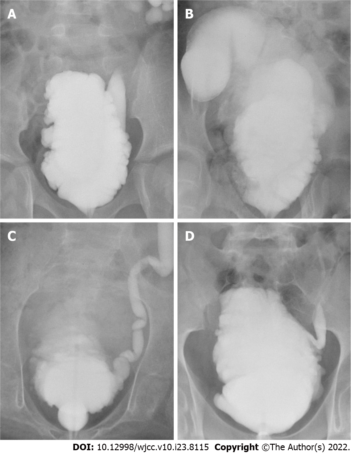
Figure 2 Cystography before and after augmentation.
A-D: the preoperative and postoperative cystography of standard cystoplasty and seromuscular bladder augmentation, respectively.
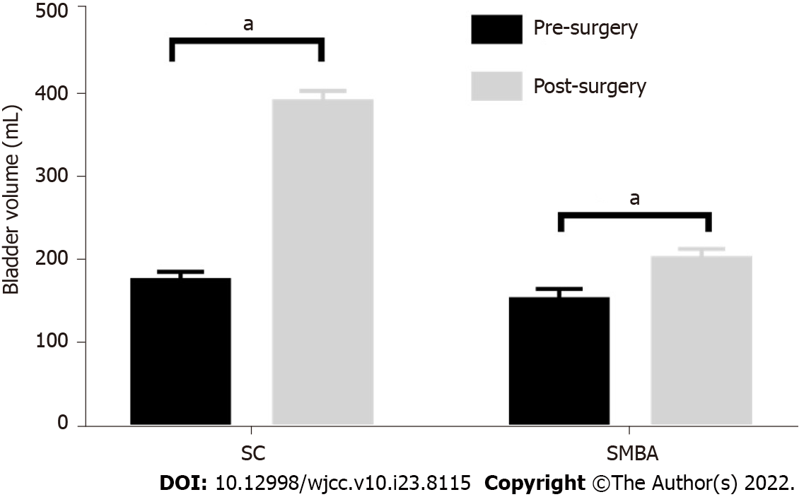
Figure 3 The preoperative and postoperative bladder volumes in the two groups were compared.
The postoperative bladder volume of the two groups was significantly enlarged compared with the preoperative volume. aP < 0.0001. SC: Standard cystoplasty; SMBA: Seromuscular bladder augmentation.
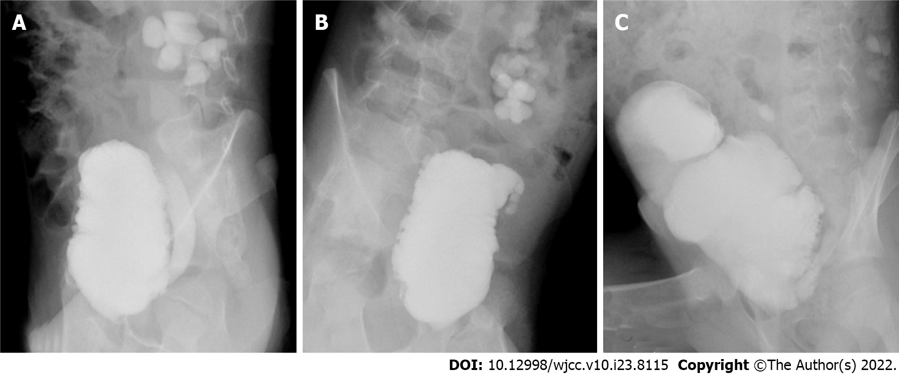
Figure 4 Reaugmentation in children with failed seromuscular bladder augmentation.
A: The neurogenic bladder before surgery; B: Patch contracture after seromuscular bladder augmentation: C: Reaugmentation with standard cystoplasty.
- Citation: Sun XG, Li YX, Ji LF, Xu JL, Chen WX, Wang RY. Outcomes of seromuscular bladder augmentation compared with standard bladder augmentation in the treatment of children with neurogenic bladder. World J Clin Cases 2022; 10(23): 8115-8123
- URL: https://www.wjgnet.com/2307-8960/full/v10/i23/8115.htm
- DOI: https://dx.doi.org/10.12998/wjcc.v10.i23.8115


