Copyright
©The Author(s) 2022.
World J Clin Cases. Jul 26, 2022; 10(21): 7609-7616
Published online Jul 26, 2022. doi: 10.12998/wjcc.v10.i21.7609
Published online Jul 26, 2022. doi: 10.12998/wjcc.v10.i21.7609
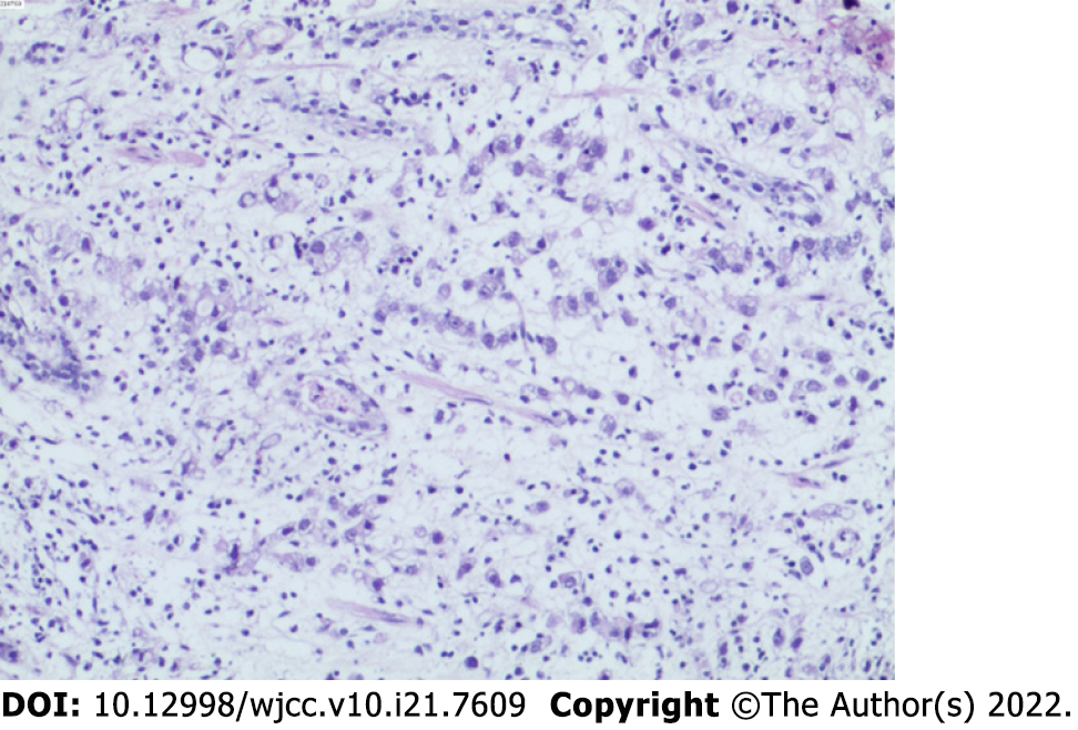
Figure 1 Biopsy of the gastric body.
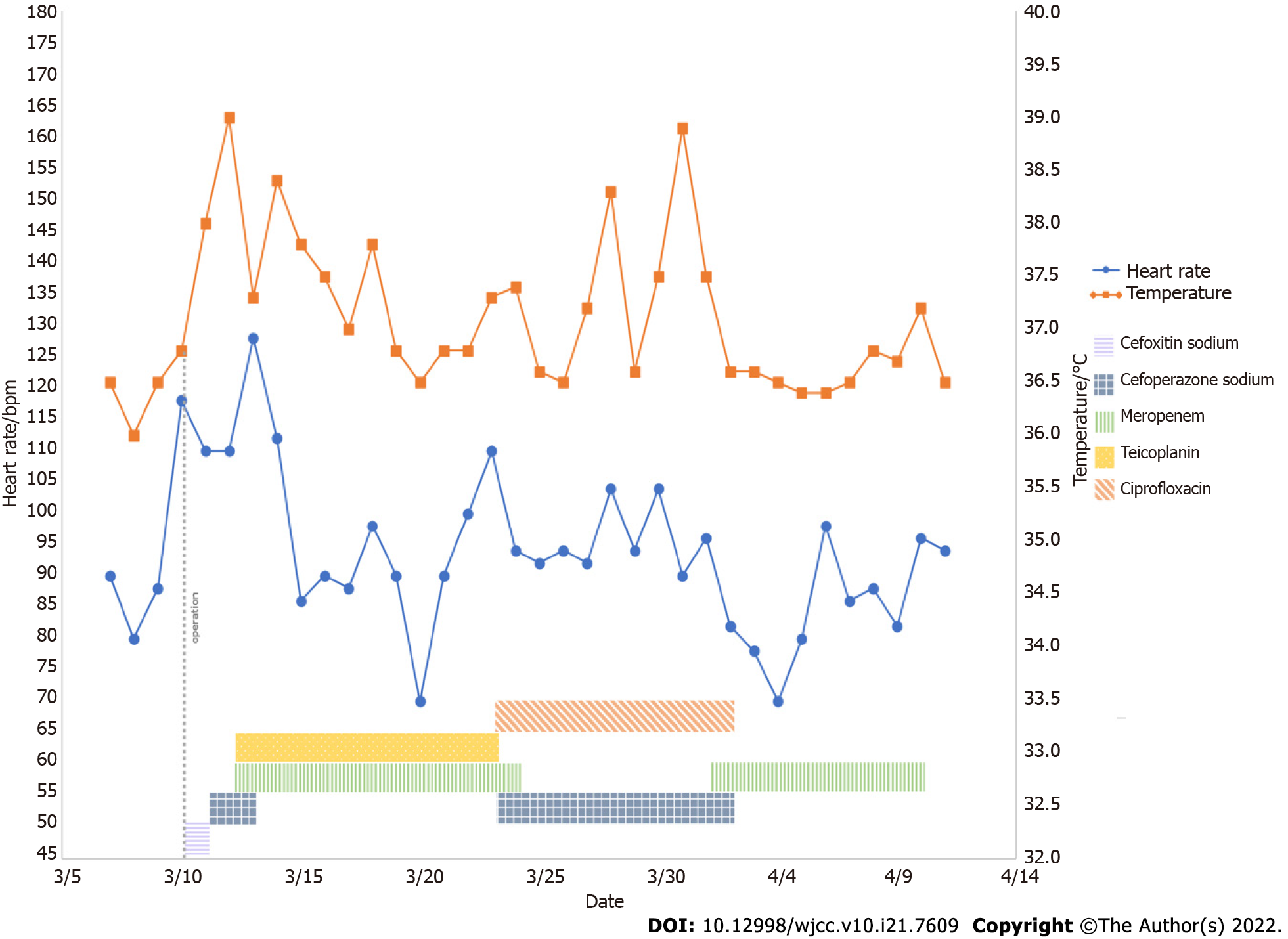
Figure 2 Basic information of the patients and the application of antibiotics during treatment.
bpm, beats per minutes.
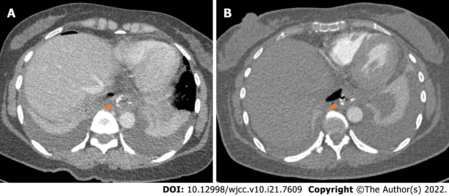
Figure 3 Postoperative anastomosis computed tomography image.
A: Computed tomography (CT) on postoperative day 6 showed changes consistent with postoperative gastrointestinal tract. The red arrow showed the position of the anastomosis; B: CT on postoperative day 13 showed anastomosis at the lower end of the esophagus. Red arrow showed the cystic air-containing cavity in the right mediastinum appears to be connected to the anastomosis and the possibility of an anastomotic fistula is considered.
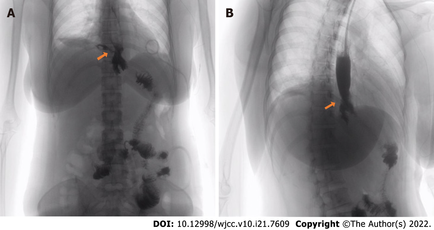
Figure 4 Radiography of gastrografin swallow.
A: Radiography of gastrografin swallow before the anastomotic fistula repair. The lower esophagus was anastomosed with jejunum after radical total gastrectomy for gastric cancer and contrast agent leakage was seen at the upper end of anastomosis; B: Radiography of gastrografin swallow after the anastomotic fistula repair. Arrow shows contrast agent leakage.
- Citation: Lu CY, Liu YL, Liu KJ, Xu S, Yao HL, Li L, Guo ZS. Differences in examination results of small anastomotic fistula after radical gastrectomy with afterward treatments: A case report . World J Clin Cases 2022; 10(21): 7609-7616
- URL: https://www.wjgnet.com/2307-8960/full/v10/i21/7609.htm
- DOI: https://dx.doi.org/10.12998/wjcc.v10.i21.7609


