Copyright
©The Author(s) 2022.
World J Clin Cases. Jul 26, 2022; 10(21): 7531-7538
Published online Jul 26, 2022. doi: 10.12998/wjcc.v10.i21.7531
Published online Jul 26, 2022. doi: 10.12998/wjcc.v10.i21.7531
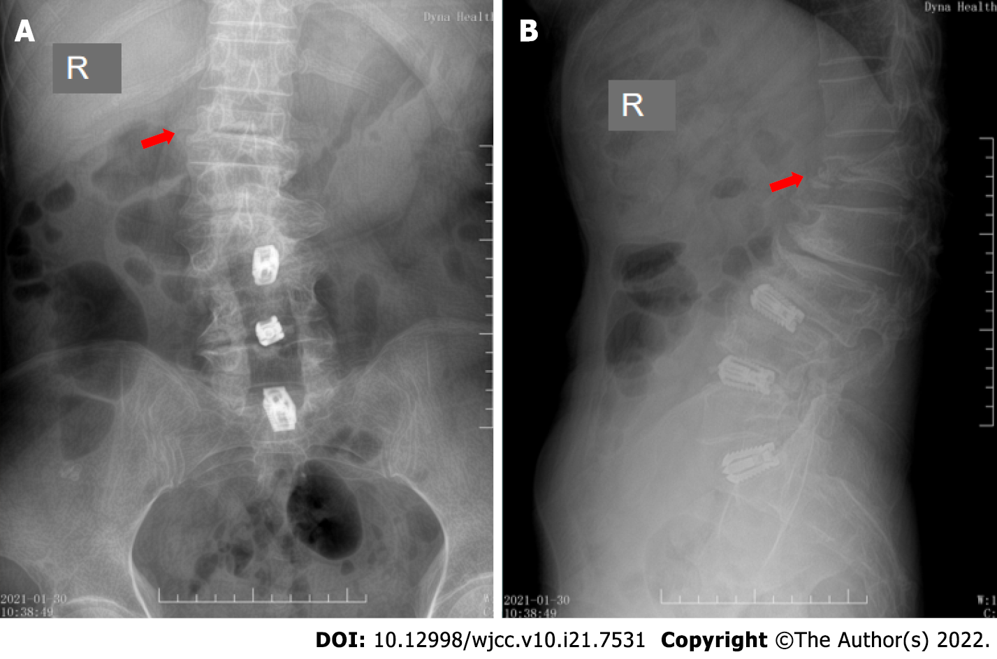
Figure 1 Preoperative X-ray image.
An osteoporotic vertebral compression fractures of the first lumbar vertebra, showing that the height of the anterior edge of the compressed vertebral body was in compression. The red arrows indicate the fractured lumbar vertebra. A: Preoperative anterior position X-ray image; B: Preoperative lateral position X-ray image. R: Right direction.
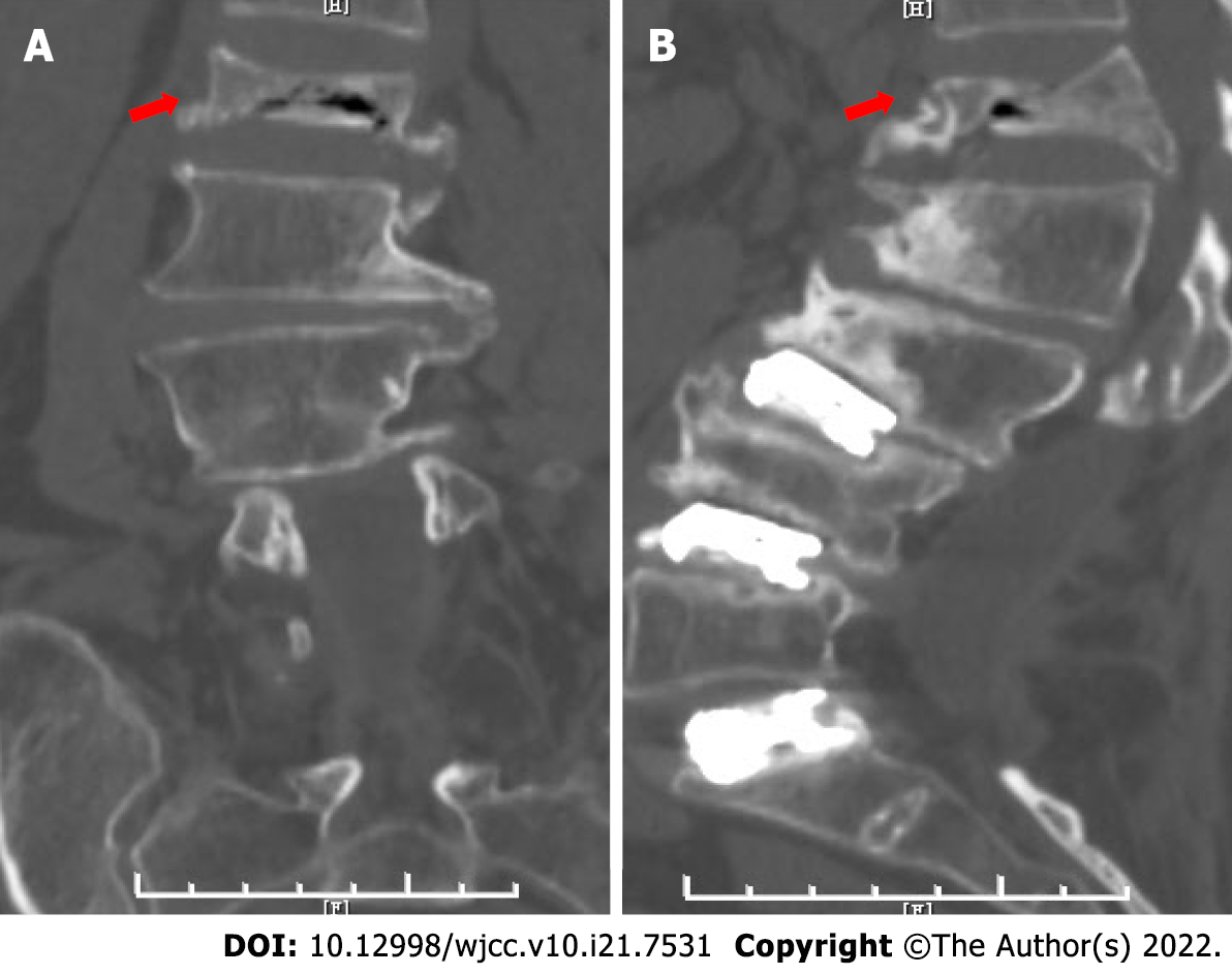
Figure 2 Preoperative computed tomography image.
The first lumbar vertebra body was compressed and there was a bone block behind the vertebral body, which compressed the spinal cord. The red arrows indicate the fractured lumbar vertebra. A: Preoperative anterior position computed tomography (CT) image; B: Preoperative lateral position CT image.
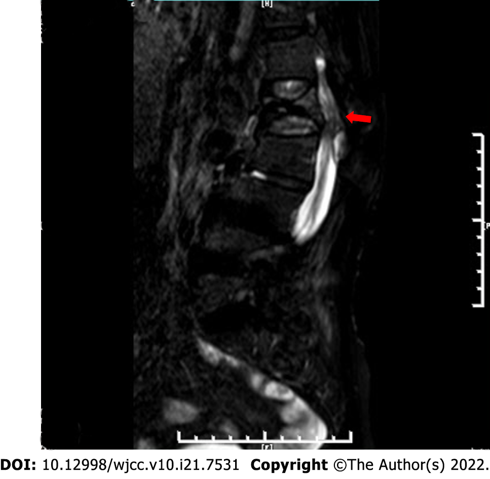
Figure 3 Preoperative magnetic resonance imaging image.
The first lumbar vertebral body was compressed, and the spinal cord was also compressed by a bone block of the fractured vertebral body. The red arrow indicates the fractured lumbar vertebra.
Figure 4 Traditional Chinese medicine manipulative reduction.
One assistant held the patient firmly from the armpit, while the other assistant held the patient’s ankles to provide fixation. At the same time, the surgeon put his hands on the spinous process of the fractured vertebrae on the patient’s back, and used it as a fulcrum to apply a posterior-anterior reduction force.
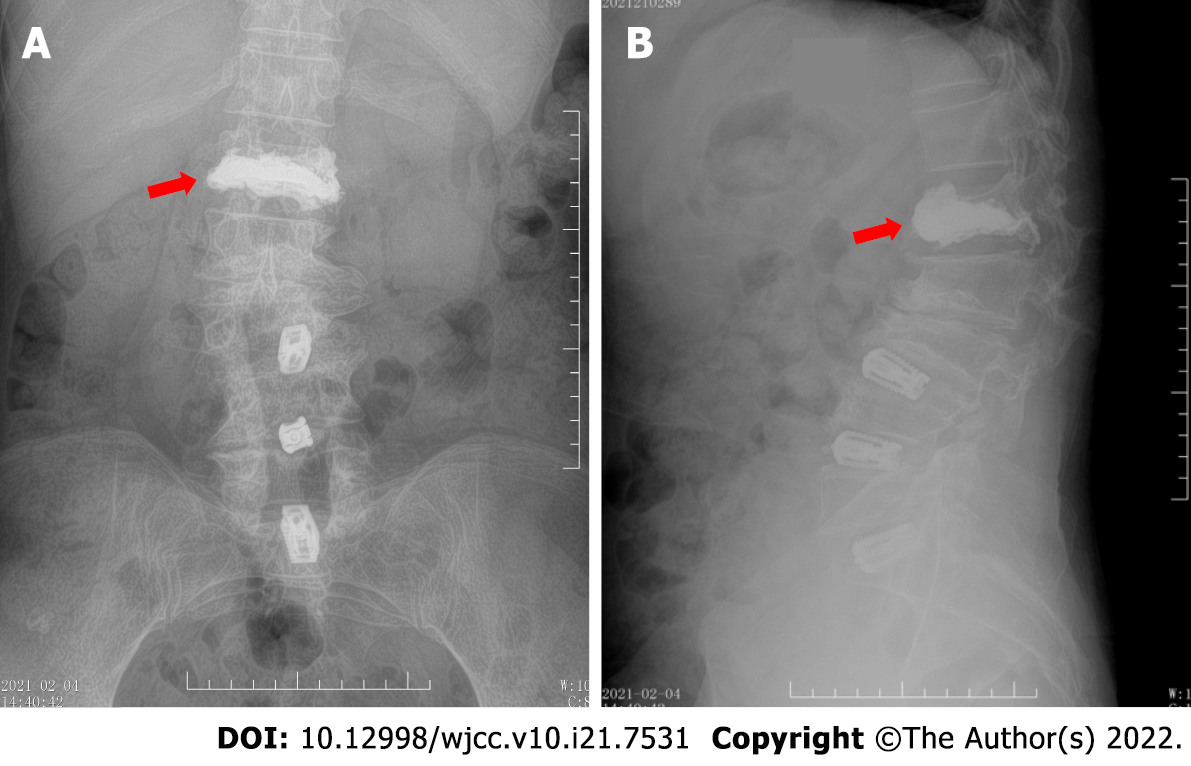
Figure 5 Postoperative X-ray image.
The height of the compressed anterior edge of the first lumbar vertebral body was reduced. The red arrows indicate the fractured lumbar vertebra, filled with bone cement. A: Postoperative anterior position X-ray image; B: Postoperative lateral position X-ray image.
- Citation: Hao SS, Zhang RJ, Dong SL, Li HK, Liu S, Li RF, Ren HH, Zhang LY. Traditional Chinese medicine manipulative reduction combined with percutaneous vertebroplasty for treating type III Kummell's disease: A case report. World J Clin Cases 2022; 10(21): 7531-7538
- URL: https://www.wjgnet.com/2307-8960/full/v10/i21/7531.htm
- DOI: https://dx.doi.org/10.12998/wjcc.v10.i21.7531


