Copyright
©The Author(s) 2022.
World J Clin Cases. Jul 26, 2022; 10(21): 7502-7508
Published online Jul 26, 2022. doi: 10.12998/wjcc.v10.i21.7502
Published online Jul 26, 2022. doi: 10.12998/wjcc.v10.i21.7502
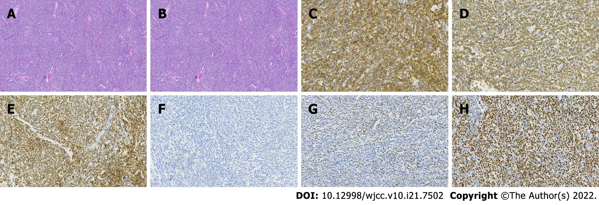
Figure 1 Histopathological microphotograph of primary testicular diffuse large B-cell lymphoma.
A: The basic structure of the testicle is destroyed, and there are a lot of lymphocytes infiltrating (magnification × 100); B: Hematoxylin and eosin stained sections showed diffuse proliferation of medium-sized round cells (magnification × 400); C-H: On immunohistochemistry, the neoplastic cells showed positive expression of CD20 (C), CD79a (D), BCL-2 (E), BCL-6 (F), C-myc (G); Ki67 staining showed almost 80% proliferation index (H).
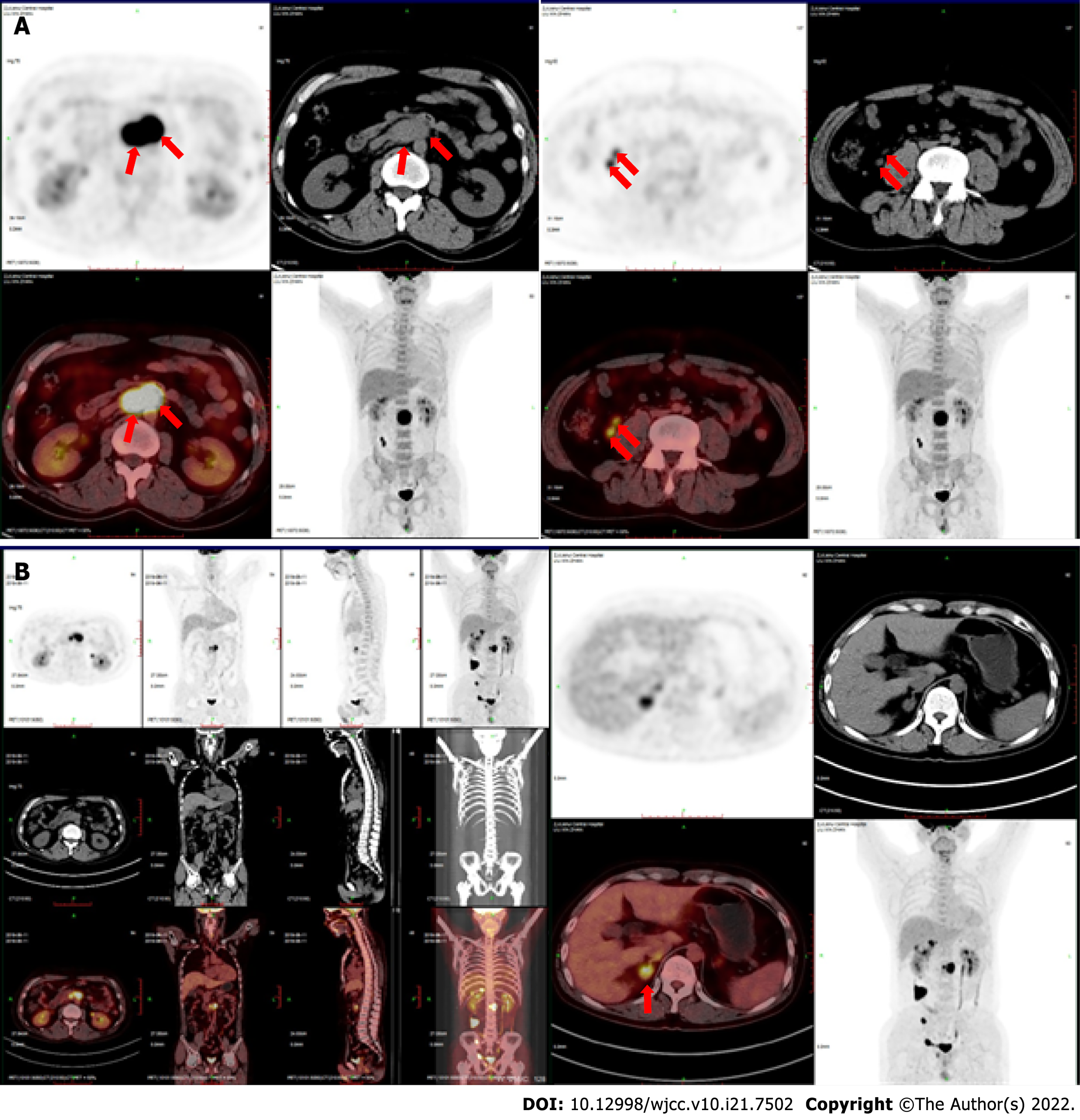
Figure 2 Positron emission tomography/computed tomography scan.
A: Positron emission tomography/computed tomography (PET-CT) scan showed multiple enlarged lymph nodes (red arrows); B: Follow-up PET-CT scan showed that, beside the right iliac vessels, the lymph nodes continued to enlarge. New viable lesions were found in the right adrenal gland, right seminal vesicle gland and surrounding prostate gland, and right groin.
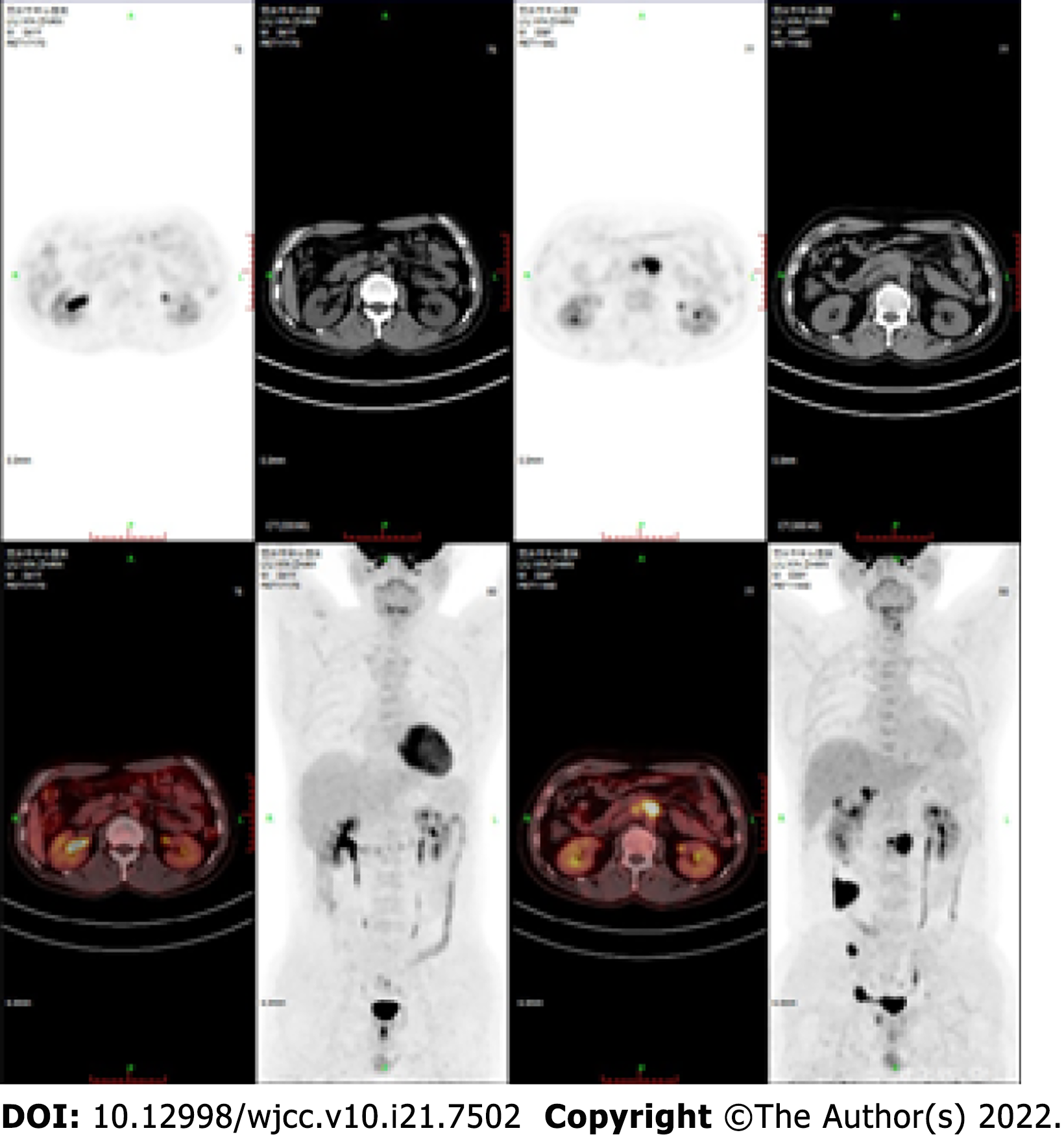
Figure 3 After chimeric antigen receptor T cells combined with programmed cell death protein-1 inhibitor treatment, positron emission tomography/computed tomography scan showed no viable lesions.
- Citation: Zhang CJ, Zhang JY, Li LJ, Xu NW. Refractory lymphoma treated with chimeric antigen receptor T cells combined with programmed cell death-1 inhibitor: A case report. World J Clin Cases 2022; 10(21): 7502-7508
- URL: https://www.wjgnet.com/2307-8960/full/v10/i21/7502.htm
- DOI: https://dx.doi.org/10.12998/wjcc.v10.i21.7502


