Copyright
©The Author(s) 2022.
World J Clin Cases. Jul 26, 2022; 10(21): 7474-7482
Published online Jul 26, 2022. doi: 10.12998/wjcc.v10.i21.7474
Published online Jul 26, 2022. doi: 10.12998/wjcc.v10.i21.7474
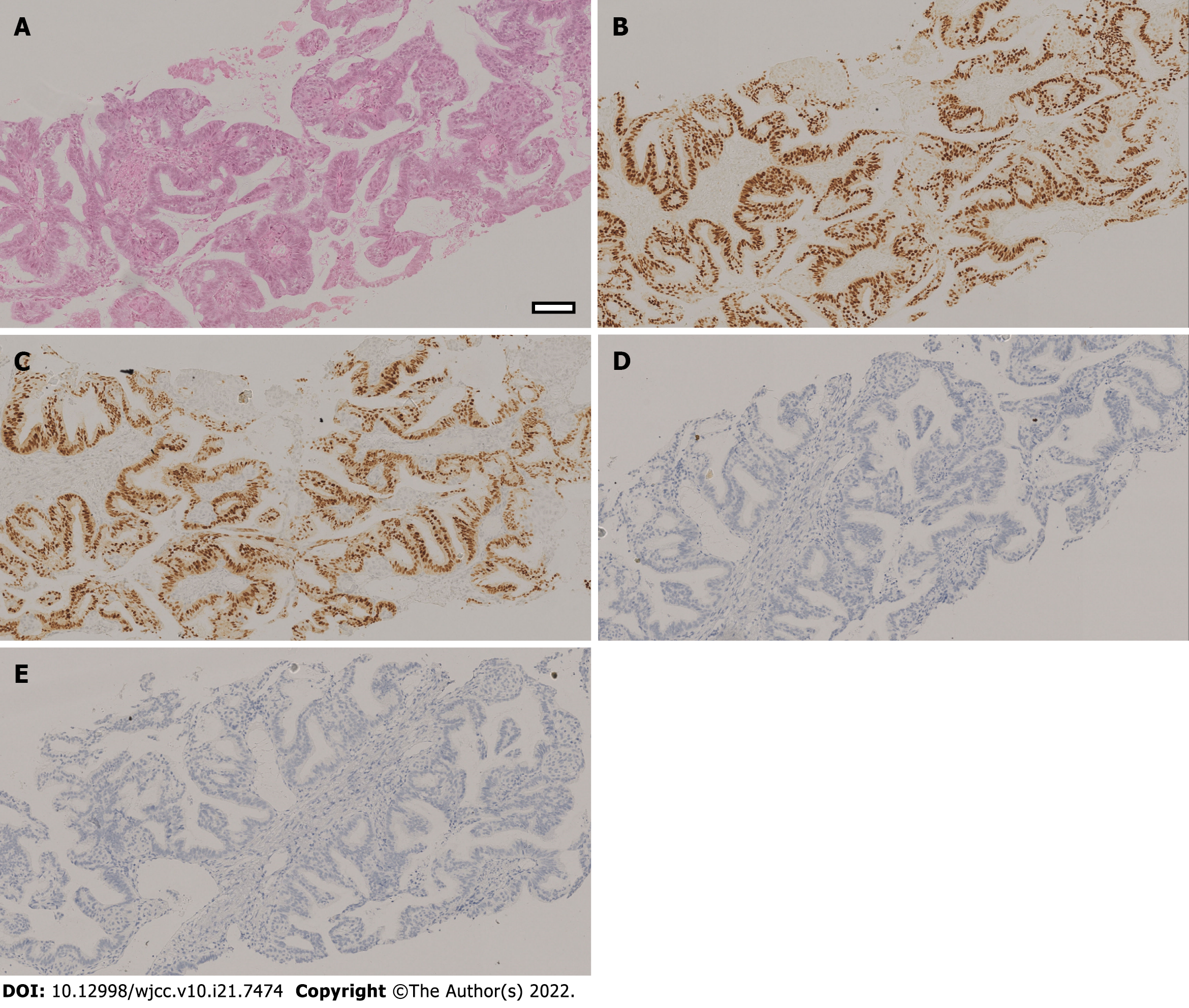
Figure 1 Pathological examination of pulmonary biopsy of the patient.
The Hematoxylin-eosin staining and immunohistochemistry staining results indicated that the endometrioid adenocarcinoma had metastasized to the lung. A: A hematoxylin and eosin-stained slide showed the glands are arranged back-to-back and crowded, with complex branches. Parts of them form papillary structures into the glandular lumen, with only a small amount of stroma. The glandular epithelium is stratified with no polar orientation. The nuclei tend to be round. The nuclear to cytoplasmic ratio was increased, the chromatin were vacuolated, and nucleoli could be seen; B: Estrogen receptor staining showing diffuse estrogen receptor expression; C: Progesterone receptor staining showing diffuse nuclear progesterone receptor expression; D: Thyroid transcription factor-1 (TTF-1) immunohistochemistry showing negative TTF-1 staining in the nucleus; E: Napsin A immunohistochemistry showing negative Napsin A staining in the cytoplasm. Original magnification: 100 ×; scale bar: 100 μm.
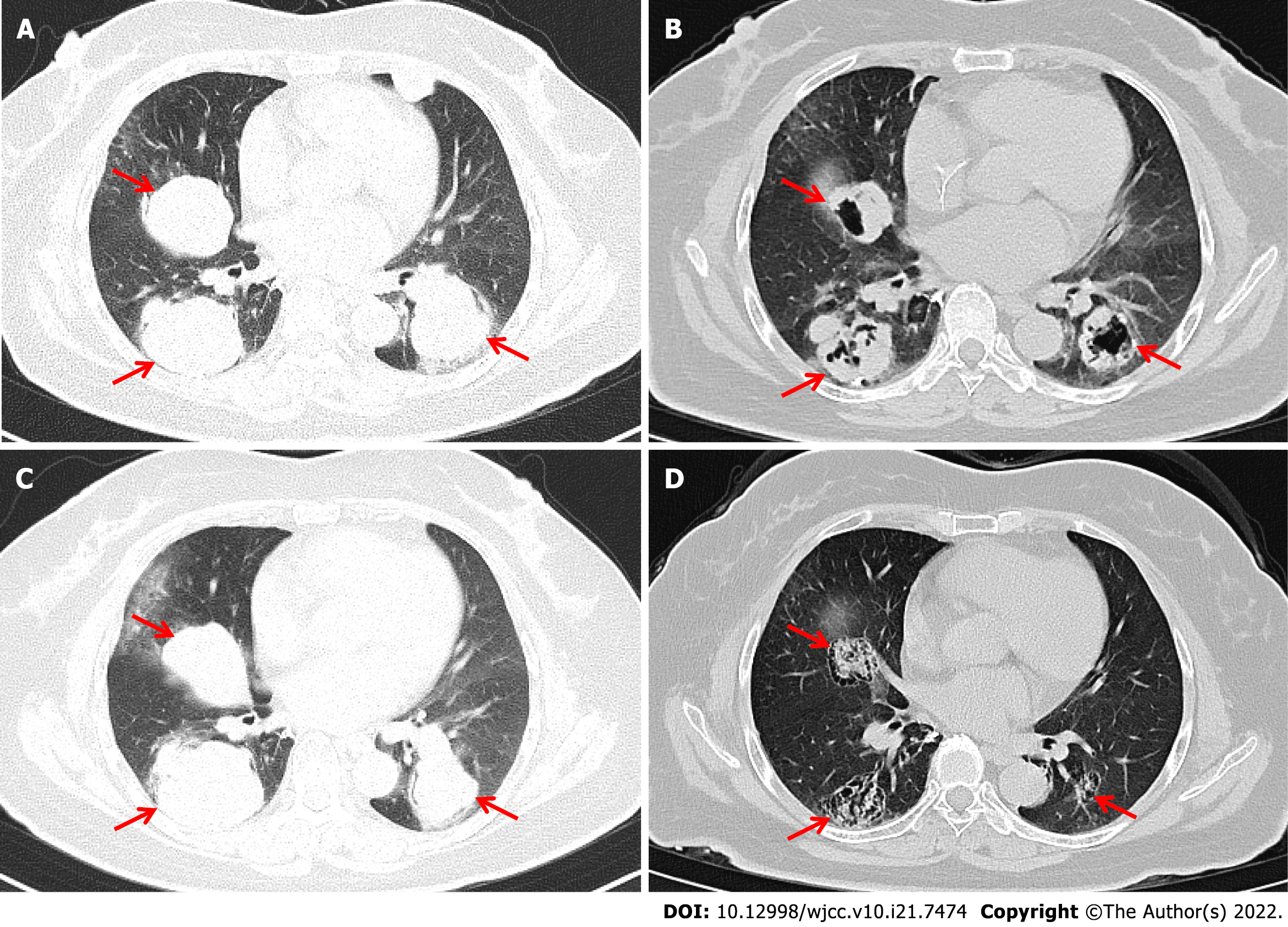
Figure 2 Computed tomography images scans showing changes in lung tumors after combined programmed cell death receptor-1 inhibitor therapy.
Chest computed tomography images showing multiple metastases in bilateral lungs. The three metastases with the largest diameters were selected as target lesions (red arrow). A: The sizes of target lesions before PD-1 inhibitor combination treatment were 47.4 mm, 50.1 mm, and 48.4 mm (6/26/2019); B: The target lesions had regressed in size to 28.9 mm, 37.2 mm, and 32.0 mm after chemotherapy combined with immunotherapy (11/28/2019); C: The target lesions were 57.6 mm, 56.9 mm, and 51.2 mm after 4.4 mo discontinuing treatment (4/16/2020); D: Target lesions had regressed in size to 26.8 mm, 35.8 mm, and 24.3 mm after immunotherapy combined with anti-angiogenesis therapy and massive necrosis was clearly observed (12/23/2020).
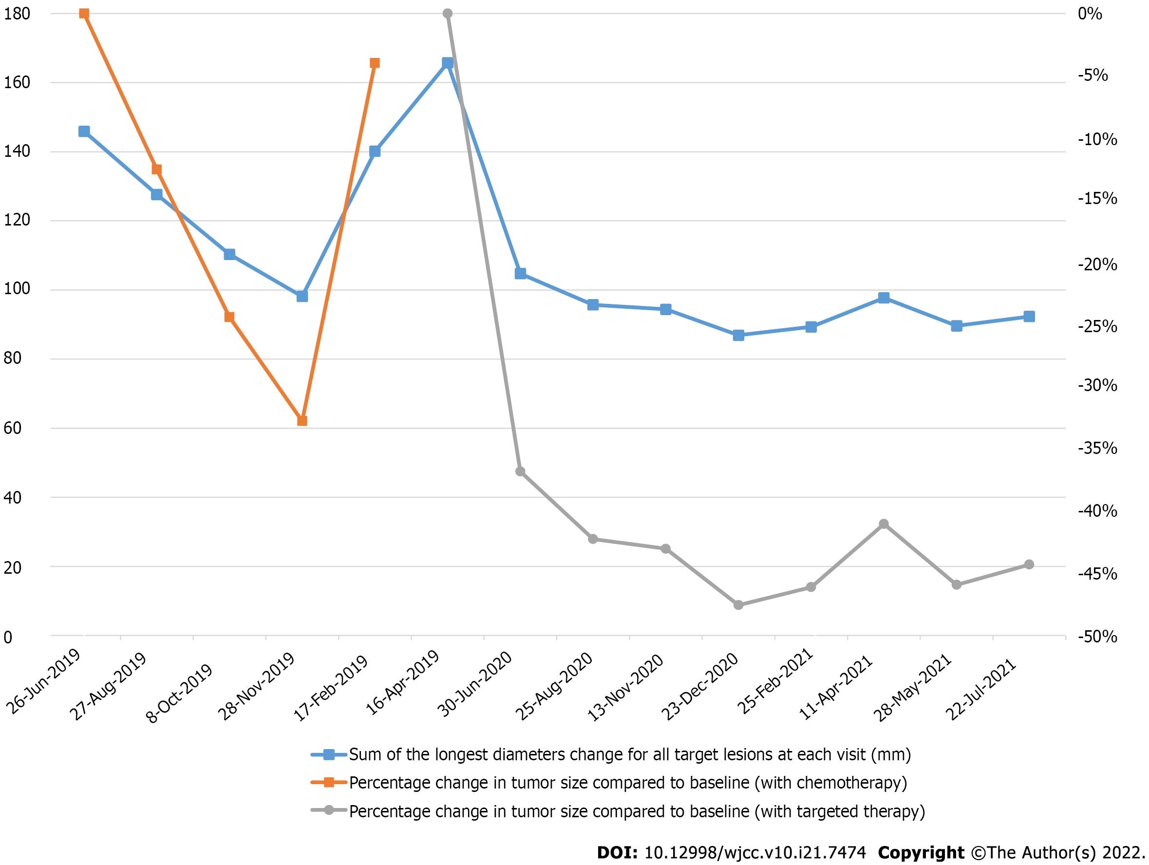
Figure 3 Sum of all longest diameters for all target lesions recorded at each visit and its percentage change from baseline during programmed cell death receptor-1 inhibitor with chemotherapy or targeted therapy.
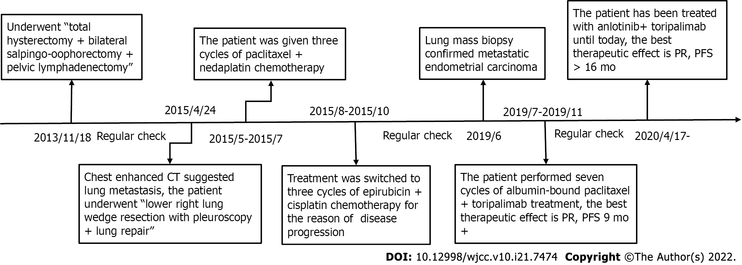
Figure 4 Timeline of diagnosis and treatment for mismatch repair proficient / microsatellite stability/human leukocyte antigen-1 heterozygous endometrial carcinoma.
- Citation: Zhai CY, Yin LX, Han WD. Programmed cell death-1 inhibitor combination treatment for recurrent proficient mismatch repair/ miscrosatellite-stable type endometrial cancer: A case report. World J Clin Cases 2022; 10(21): 7474-7482
- URL: https://www.wjgnet.com/2307-8960/full/v10/i21/7474.htm
- DOI: https://dx.doi.org/10.12998/wjcc.v10.i21.7474


